Diagnostic Tests for Neurological Disorders
Diagnostic Tests for Neurological Disorders
What are some diagnostic tests for nervous system disorders?
In addition to a complete medical history and physical exam, diagnostic procedures for nervous system disorders may include the following:
Computed tomography scan (also called a CT or CAT scan). A diagnostic imaging procedure that uses a combination of X-rays and computer technology to produce horizontal, or axial, images of the body. A CT scan shows detailed images of any part of the body, including the bones, muscles, fat, and organs. CT scans are more detailed than general X-rays.
Electroencephalogram (EEG). A procedure that records the brain's continuous electrical activity through electrodes attached to the scalp.
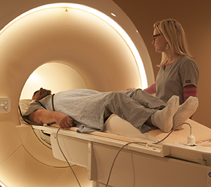
Magnetic resonance imaging (MRI). A diagnostic procedure that uses a combination of large magnets, radiofrequencies, and a computer to produce detailed images of organs and structures within the body.
Electrodiagnostic tests, such as electromyography (EMG) and nerve conduction velocity (NCV). Studies that evaluate and diagnose disorders of the muscles and motor neurons. Electrodes are inserted into the muscle, or placed on the skin overlying a muscle or muscle group, and electrical activity and muscle response are recorded.
Positron emission tomography (PET). In nuclear medicine, a procedure that measures the metabolic activity of cells.
Arteriogram (also called an angiogram). An X-ray of the arteries and veins to detect blockage or narrowing of the vessels.
Spinal tap (also called a lumbar puncture). A special needle is placed into the lower back, into the spinal canal. This is the area around the spinal cord. The pressure in the spinal canal and brain can then be measured. A small amount of cerebral spinal fluid (CSF) can be removed and sent for testing to determine if there is an infection or other problems. CSF is the fluid that bathes the brain and spinal cord.
Evoked potentials. Procedures that record the brain's electrical response to visual, auditory, and sensory stimuli.
Myelogram. A procedure that uses dye injected into the spinal canal to make the structure clearly visible on X-rays.
Neurosonography. A procedure that uses ultra high-frequency sound waves that enable the healthcare provider to analyze blood flow in cases of possible stroke.
Ultrasound (also called sonography). A diagnostic imaging technique that uses high-frequency sound waves and a computer to create images of blood vessels, tissues, and organs. Ultrasounds are used to view internal organs as they function, and to assess blood flow through various vessels.
Updated:
April 25, 2018
Sources:
Overview of Electromyography, Up To Date
Reviewed By:
Shelat, Amit, MD,Turley, Raymond Kent, BSN, MSN, RN