Diagnosing Chest and Lung Problems: Imaging Tests
Diagnosing Chest and Lung Problems: Imaging Tests
CT scan
CT scans allow the healthcare provider to see a more detailed picture of the chest and lungs than a regular chest X-ray. During a CT scan, many images are taken of the lungs and chest. A computer combines the images to create one detailed image. In some cases, special dye (contrast) is given through an IV (intravenous) line. The contrast highlights any suspicious area on the scan.
PET scan
Positron emission tomography (PET) is used to diagnose chest and lung problems. For a PET scan, a safe radioactive liquid (tracer) is injected into the bloodstream. It takes about 45 minutes for the tracer to be in your system. Once the tracer is there, the healthcare provider takes a scan of your body. A PET scan can be helpful for finding cancer. It can also help find out if a cancer has spread. This is called staging. In some cases, you may need more testing before cancer is diagnosed or ruled out.
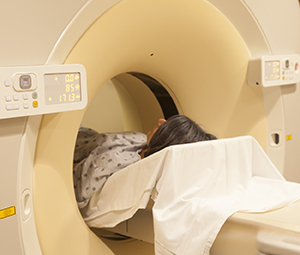
MRI
Like the CT scan, MRI takes many detailed images of the chest and lungs without the use of X-ray radiation. Contrast dye may be given through an IV line. The healthcare provider then takes a scan. MRI helps your provider find out if a mass is affecting other structures in the chest, such as blood vessels.
Getting ready for the test
Before your imaging test, do the following:
Follow your healthcare provider’s instructions about eating and drinking
Tell your provider about the medicines you take. You may need to stop taking certain medicines before the test.
Discuss any allergies and health problems with your healthcare provider. Be sure to tell him or her if you are allergic to iodine or contrast or if you have kidney problems.
Tell your healthcare provider and the healthcare person doing the scan if you wear a medicated adhesive patch.
Mention if you have any metal in your body. This includes loose pieces of metal or metal devices such as an aneurysm clip, a pacemaker, a prosthesis, braces on your teeth, or an intraocular lens.
Tell your healthcare provider if you are pregnant.
During the test
For the test, you lie on your back inside a tube-like machine (scanner). You hear loud clicking sounds as images are taken. To make sure the images taken are clear, you must lie still. Straps may be used to help with this. Imaging tests (especially MRI) are done in a confined space. Talk with your healthcare provider before the day of your test if you are afraid of confined spaces. You may be given medicine (sedation) to help you relax during the test.
Risks and complications
Swelling, infection, or other problems at the IV site
Contrast or tracer-related problems, such as allergic reaction or kidney damage
Damage to metal devices or prostheses from large magnetic MRI scanner
Updated:
December 22, 2017
Reviewed By:
Adler, Liora C., MD,Sather, Rita, RN,Image reviewed by StayWell art team.