Hip Arthroscopy: Repairing Femoroacetabular Impingement
Hip Arthroscopy: Repairing Femoroacetabular Impingement
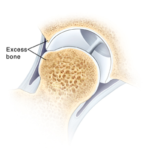
When excess bone has formed on the edge of the “ball” (femoral head) or the “socket” (acetabulum) of the hip, it is called femoroacetabular impingement (FAI). FAI can cause pain and limit movement. Arthroscopy can repair FAI with only small incisions and special instruments.
In the operating room
Just before surgery, you may be asked several times which hip is to be treated. This is a standard safety measure. In the operating room, you will likely receive general anesthesia to make you sleep.
During the procedure
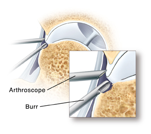
After you receive anesthesia, your leg is gently pulled to distract, or widen, the hip joint. Next, the surgeon makes a few small incisions called portals. Through these portals, he or she inserts surgical tools, including the arthroscope. The arthroscope sends images of the joint to a video screen. These images allow the surgeon to look inside the joint. The joint is filled with sterile fluid to help the surgeon see more clearly.
Repairing femoroacetabular impingement
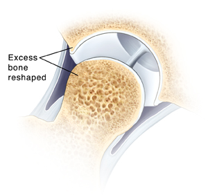
To treat FAI, the area is reshaped by removing excess bone. Excess bone can be removed from the socket side or ball side of the hip joint, or both. FAI can lead to other problems, such as labral tears or chondral damage. If present, these problems are also treated. Once the surgeon finishes the procedure, the portals are closed and bandaged. Then you are taken to the recovery room.
Updated:
March 21, 2017
Reviewed By:
Image reviewed by StayWell medical illustration team.,Joseph, Thomas N., MD,Moloney Johns, Amanda, PA-C, MPAS, BBA