Craniotomy: Correcting Your Problem
Craniotomy: Correcting Your Problem
Craniotomy is a surgical opening made in the skull for treatment of several types of problems in the brain. Special tools are used to remove a piece of the skull and allow access to the brain for surgical treatment. The most commons reasons for having a craniotomy include a blood clot (hematoma), tumors, aneurysms, arteriovenous malformations (AVM), and brain abscess.
What your surgeon does during your craniotomy depends on your problem. But no matter what, every measure is taken to avoid damage to normal tissue.
Brain injuryThe source of bleeding is controlled and blood is removed. Damaged tissue may also be cleaned away. |
Brain tumorAs much of the brain tumor as safely possible is removed. |
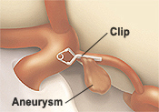
AneurysmThe artery is clipped or sealed at the leak. This prevents more blood from flowing around or into the brain. |
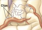
AVM (arteriovenous malformation)Abnormal arteries and veins are clipped to redirect blood flow to normal vessels and prevent the AVM from leaking blood. After the abnormal arteries and veins are sealed off, they may be removed through the blood vessels using a glue-like substance. This process is called embolization and resection. |

Other types of brain procedures
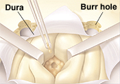
The procedures below may be done alone, or they may be done in addition to a craniotomy. To provide access for shunts or stereotactic surgery, burr holes are made in the skull. Your hospital experience before and after these procedures may be about the same as for a craniotomy.
Shunts. A shunt is a special type of drain. It is used to decrease pressure on the brain by removing excess cerebrospinal fluid (CSF). The fluid is drained from the brain to the abdomen or another cavity of the body. This is done through a tube that is tunneled under the skin. Once the fluid reaches the abdomen or another cavity, the body absorbs it. Sometimes the CSF drain is temporarily (usually for a few days) connected to an external collection bag, this is called an external ventricular drain (EVD).
Brain mapping. The surgeon may monitor responses during the operation to identify regions with important functions.
Updated:
April 27, 2018
Reviewed By:
Jasmin, Luc, MD,Sather, Rita, RN