Heart rate variability
Heart rate variability
Natural Standard Monograph, Copyright © 2013 (www.naturalstandard.com). Commercial distribution prohibited. This monograph is intended for informational purposes only, and should not be interpreted as specific medical advice. You should consult with a qualified healthcare provider before making decisions about therapies and/or health conditions.
Related Terms
Arrhythmia, atrial fibrillation, autonomic nervous system (ANS), Bainbridge reflex, beat-to-beat interval, bradycardia, cardiac arrest, cardiac event, central nervous system (CNS), circadian rhythm, cycle length variability, deceleration capacity, digital photoplethysmography, electrocardiogram (ECG or EKG), electrocardiography, Electrokardiogramm (German), expiratory-to-inspiratory ratio (E:I), fingertip photoplethysmography, Fourier transformation, Framingham Heart Study, frequency domain, heart attack, heart period variability, heart rate, heart rate variability, heart rate variability with deep breathing (HRVdb), heart rate variation, implantable cardioverter-defibrillator (ICD), mean heart rate range (MHRR), myocardial infarction (MI), normal-to-normal (NN) interval, P wave, parasympathetic nervous system, peripheral nervous system (PNS), photoplethysmography (PPG), plethysmography, QRS complex, RR interval, RR period, RR variability, sudden cardiac death, sympathetic nervous system, T wave, tachycardia, time domain, U wave, ventricular fibrillation.
Background
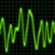
Heart rate variability (HRV) is defined as changes in the duration of consecutive cardiac cycles (heartbeats). Cardiac cycles may be measured by electrocardiography (ECG), which produces an electrocardiogram (EKG, for "Electrokardiogramm" in German). On an EKG, the pattern of a cardiac cycle has four major parts: a P wave (which represents the electrical vector spreading from the right atrium to the left atrium during atrial depolarization), a QRS complex (which represents depolarization of the right and left ventricles), a T wave (which represents repolarization of the ventricles), and a U wave (which represents repolarization of the papillary muscles). HRV is measured as the variation in duration between the R peaks on the QRS complexes between consecutive cardiac cycles. Other names for HRV include RR variability, RR period, cycle length variability, and heart period variability.
The autonomic nervous system (ANS) controls heart rate, HRV, and other involuntary functions such as breathing. HRV reflects the complex autonomic controls of heart rate. Thus, HRV measurements contain more information than measurements of heart rate alone.
HRV was first described in 1847, when respiration (breathing) was noted to affect HRV by increasing heart rate during inspiration (breathing in) and decreasing during expiration (breathing out). The clinical significance of HRV was realized in 1963, when researchers showed that just before fetal death, changes in beat-to-beat length were detectable before changes in the heart rate itself.
In 1987, HRV was reported to have prognostic value when decreased HRV was found to be associated with decreased survival (increased death or mortality) in myocardial infarction (MI or heart attack) patients. Because numerous studies have demonstrated a link between HRV and poor prognosis after MI, HRV measurements are widely accepted for predicting risk of future cardiac events (heart problems) in heart attack survivors.
HRV is also strongly associated with arrhythmias (abnormal heart rate), sudden cardiac death, and all fatalities (all-cause mortality) in cardiovascular diseases in general. Sudden cardiac death due to ventricular arrhythmia is a leading cause of death in the United States, accounting for up to 500,000 deaths each year. Because measures of HRV are easily obtained and non-invasive, HRV is gaining favor as a method of assessing autonomic function in routine clinical practice.
HRV has been used to screen candidates for implantable cardioverter-defibrillator (ICD) devices that may help control arrhythmias and prevent sudden cardiac death. However, the usefulness of HRV in screening ICD candidates has not been shown clearly. Though most of the findings thus far are promising, additional evidence is needed to support the use of HRV (alone or with other cardiac risk measurements) in predicting sudden cardiac death in various cardiac conditions. HRV may also be altered in numerous non-cardiovascular conditions and neuropathies (nerve disorder). Thus, HRV has been suggested to be useful for assessing risk for a number of cardiovascular and neurological diseases. Improving the diagnostic and prognostic value of HRV may require further optimizing its measurement techniques for specific conditions.
Contributing Factors
General: Heart rate variability (HRV) is controlled by the autonomic nervous system (ANS), also known as the visceral nervous system. The ANS is part of the peripheral nervous system (PNS). The nerves in the PNS connect various organ systems to the central nervous system (CNS), which includes the brain and spinal cord. The ANS functions at the subconscious level. In addition to controlling heart rate, it controls bodily functions such as sweating, digestion, and breathing. Because the autonomic control of heart rate can reveal abnormal changes in physiological functions, HRV may be very useful in making diagnostic, prognostic, and therapeutic determinations.
In addition to pathological (disease) conditions, there are a number of physiological factors in healthy individuals that may affect heart rate variability (HRV). While some of these factors (such as age and genetic makeup) cannot be controlled by individuals, other factors such as lifestyle choices (such as physical activity, smoking, and other lifestyle choices) are modifiable. Therefore, many lifestyle choices may affect HRV and its role in disease outcome.
Physiological factors:
Breathing: Respiration (breathing) is known to affect HRV. While it is not entirely clear exactly how breathing affects HRV, evidence from research conducted in canines suggests that an autonomous reflex known as the Bainbridge reflex may be involved. Cardiac (heart) reflexes and respiratory activities (such as rib cage movement) are controlled by the central nervous system (CNS), and thus may also affect HRV.
Circadian rhythm: The effects of circadian rhythm on HRV have been studied in healthy men and women. HRV varies over a 24-hour period, peaking at night and plateauing during the day.
Posture: In healthy subjects, rising from a supine (lying down) position to the upright (standing up) position increases resting heart rate and decreases the frequency of HRV. In the European Project on Genes in Hypertension (EPOGH) study, HRV was found to consistently vary according to posture, independently of other factors.
Non-modifiable factors:
Age: It has been firmly established that maximal heart rate becomes lower as individuals grow older. Age is known to affect autonomic control of the cardiovascular system, and is a primary factor that may affect HRV. In the European Project on Genes in Hypertension (EPOGH) study, HRV was found to consistently vary with age.
Gender: Females, under age 30, tend to have lower HRV than age-matched males. Gender differences in HRV begin to disappear at age 30 and completely disappear by age 50. This may be because as people age, sympathetic activity tends to decline more slowly in males than females.
Genetics: HRV has been examined in the Framingham Heart Study, which is a large multigenerational cohort study that began in 1948. Genomic evidence from this study strongly suggests that heart rate and HRV characteristics may be inherited and shared over multiple generations. The genes that appear to be involved include those that control the ANS and certain neural responses (such as those mediated by the cholinergic system).
Modifiable lifestyle factors:
Physical activity: The ANS controls heart rate changes during physical activity. Regular physical activity decreases heart rate during both rest and exercise in humans. It is still not exactly clear how regular physical training affects HRV. However, in several studies conducted in canines, endurance training (treadmill running) increased HRV and lowered risk for sudden cardiac death due to arrhythmia.
Smoking: Smoking is known to harm the cardiovascular system in part by increasing heart rate and reducing HRV. This effect has also been demonstrated in humans and animals exposed to second-hand tobacco smoke, as well as in infants of smoking mothers.
Other factors:
Medications: Various medications, particularly those with anticholinergic effects (such as tricyclic antidepressants and antispasmodics), are known to reduce HRV. Moreover, stimulants (such as caffeine and nicotine) increase heart rate and decrease HRV. Atenolol (a beta-antagonistic drug) has been shown to reduce HRV while losartan (angiotensin II receptor antagonist) increases HRV.
Pollution: Small-particulate air pollution and second-hand cigarette smoke have both been shown to affect the ANS. Increased heart rate and decreased HRV are associated with an increased risk of cardiovascular disease and related death.
Technique
General: Unlike simple heart rate, which does not need sophisticated technology or significant time to measure, heart rate variability (HRV) measurements involve special devices, statistical analysis, and continuous monitoring (often for 24 hours or longer).
HRV is generally measured using electrocardiography (ECG). The electrocardiogram (EKG, for "Electrokardiogramm" in German) represents the cardiac cycle as a wave with four major parts: a P wave, a QRS complex, a T wave, and a U wave. HRV is measured as the variation in duration between the R peaks on the QRS complexes over consecutive cardiac cycles. The variability of the RR interval may be measured in various ways, usually using time or frequency domain methods. Newer methods of analysis have also been developed.
Time domain methods: The simplest measures of HRV are based on time domain methods, which determine the intervals of cardiac cycles. On an ECG recording, this is usually measured as the normal-to-normal (NN) interval between QRS complexes. The instantaneous heart rate may also be determined. Variation is then calculated as the difference in NN intervals (cycle length) or heart rate. These differences may then be calculated statistically as the standard deviation of NN intervals (SDNN). Typical recording times for statistical analysis are 24 hours, but short-term recordings of five minutes may also be used. The NN intervals may also be analyzed using geometric or fractal techniques by converting the intervals into geometric patterns. When NN interval measurements are of low quality, geometric analysis is more accurate that statistical analysis. However, because many NN intervals are needed for conversion into geometric patterns, fractal or geometric methods usually require recording times of 24 hours or more and are thus not appropriate for analyzing shorter recordings (less than 20 minutes).
Frequency domain methods: HRV may be studied in the frequency domain by converting heart rate (time domain) to a power spectrum (frequency domain) using a mathematical algorithm called the Fourier transformation. High frequencies (over 0.15 Hz) indicate respiratory sinus arrhythmia, while lower frequencies reflect autonomic (sympathetic and parasympathetic) factors. Reduced frequency occurs in patients with autonomic problems, such as diabetic neuropathy. Frequency domain measures of HRV are more difficult to perform than time domain analyses. However, for continuous recordings of longer time periods (such as 24 hours), time and frequency domain measurements are generally in agreement.
Heart rate variability with deep breathing: Deep breathing amplifies HRV, and combining HRV with deep breathing (HRVdb) is a very sensitive method of measuring ANS function. HRVdb methods correlate HRV with respiratory cycles, and usually measure the mean heart rate range (MHRR) and the expiratory-to-inspiratory ratio (E:I). HRVdb has been used reliably in autonomic function tests in various autonomic disorders, including neuropathies, neurodegenerative disorders, and autonomic failure.
Deceleration capacity: A relatively new method of analyzing heart rate dynamics uses measures of HRV associated with decreasing (decelerating) but not increasing (accelerating) heart rate. This method is thought to specifically measure cardiac vagal tone, which comprises vagus nerve impulses that inhibit heartbeat. In a blind multicenter clinical study, altered deceleration capacity was shown to be a more powerful and accurate predictor of death after myocardial infarction (MI or heart attack) than standard HRV measurements.
Photoplethysmography (PPG): The plethysmogram waveform represents pulsatile peripheral blood flow, which reflects both peripheral and central hemodynamics. PPG uses infrared light transmitted through the skin to noninvasively measure hemodynamic parameters, and is thus a useful measure of vascular dysfunction and HRV. When used to measure HRV in subjects at rest, fingertip PPG is about as accurate as ECG.
Theory/Evidence
There is a large body of evidence linking altered heart rate variability (HRV) with death, particularly in deaths due to arrhythmia. Numerous studies have demonstrated a link between HRV and poor prognosis after myocardial infarction (MI or heart attack). Therefore, HRV measurements are widely accepted in for predicting future complications and death in MI survivors.
A landmark study in 1987 tested whether HRV could predict long-term survival after MI. This study found that decreased HRV was strongly correlated with death after MI. Data reported in 1996 from the Framingham Heart Study found that in this large community-based study population, decreased HRV was associated with increased risk of major heart problems after an average of 2.5 years, even after adjusting for other known cardiovascular risk factors. The TRAndolapril Cardiac Evaluation (TRACE) study and the Autonomic Tone and Reflexes After Myocardial Infarction (ATRAMI) study each provided additional evidence that decreased HRV is linked with increased death rates after MI.
For non-ischemic cardiomyopathies, there are conflicting reports of association between reduced HRV and poor prognosis. However, in valvular heart diseases (such as chronic mitral regurgitation), lower HRV appears to predict future heart problems. There is also evidence that reduced HRV may predict postoperative cardiac complications, which causes death in many cases of postoperative cardiac arrests.
There is evidence that decreased HRV reflects autonomic disturbances, which may increase the risk of ventricular fibrillation. Decreased HRV has also been shown to correlate with increased inflammatory markers, such as interleukin-6 (IL-6) and C-reactive protein (CRP), which are associated with increased risk of adverse cardiac events. In a prospective cohort study, autonomic dysfunction (indicated by reduced HRV) and increased inflammation preceded sudden cardiac death in chronic heart failure patients.
In a study involving 202 patients with severe chronic heart failure, short-term HRV was found to be a strong predictor of sudden cardiac death. This correlation has also been demonstrated in a random sample of 325 elderly subjects, in which fractal analysis of HRV strongly predicted cardiac death over a period of 10 years.
The prognostic value of HRV for sudden cardiac death may be improved by combining HRV measure with other parameters, such as heart rate turbulence and neurohormonal activation.
HRV is also altered in a number of non-cardiac diseases, including brain damage, depression, diabetes, end-stage renal disease, epilepsy, Guillain-Barré syndrome, schizophrenia, and neuropathy. Many of these diseases carry an increased risk of sudden cardiac death. Therefore, monitoring HRV has been considered as a method for helping prevent sudden cardiac death in these conditions.
Safety
Heart rate variability (HRV) is strongly correlated with fatal arrhythmias and sudden cardiac death. Therefore, it has been used to screen candidates for implantable cardioverter-defibrillators (ICDs). Although HRV measurements are non-invasive and may provide useful prognostic information, it may not be safe to select patients for therapy based on HRV measurements alone.
In the Defibrillator in Acute Myocardial Infarction Trial (DINAMIT), altered HRV was used to select candidates for ICD placement in survivors of acute myocardial infarction (MI or heart attack). Although there were fewer deaths among ICD patients than in non-ICD patients, this decreased death was offset by more deaths from nonarrhythmic causes. In the AzimiLide post Infarct surVival Evaluation (ALIVE) trial, stratifying (ranking) patients according to HRV also did not affect death rates in patients treated with azimilide (an antiarrhythmic drug). This may be because HRV has not yet been optimized for predicting death from non-arrhythmic effects of drugs.
Author Information
This information has been edited and peer-reviewed by contributors to the Natural Standard Research Collaboration (www.naturalstandard.com).
Bibliography
Natural Standard developed the above evidence-based information based on a thorough systematic review of the available scientific articles. For comprehensive information about alternative and complementary therapies on the professional level, go to www.naturalstandard.com. Selected references are listed below.
Bauer A, Kantelhardt JW, Barthel P, et al. Deceleration capacity of heart rate as a predictor of mortality after myocardial infarction: cohort study. Lancet. 2006 May 20;367(9523):1674-81. View Abstract
Framingham Heart Study: A Project of the National Heart, Lung, and Blood Institute and Boston University. www.framinghamheartstudy.org
Gang Y, Malik M. Non-invasive risk stratification for implantable cardioverter-defibrillator placement--heart rate variability. Am Heart Hosp J. 2009 Summer;7(1):39-44. View Abstract
Giardino ND, Lehrer PM, Edelberg R. Comparison of finger plethysmograph to ECG in the measurement of heart rate variability. Psychophysiology. 2002 Mar;39(2):246-53. View Abstract
Hohnloser SH, Kuck KH, Dorian P, et al.; DINAMIT Investigators. Prophylactic use of an implantable cardioverter-defibrillator after acute myocardial infarction. N Engl J Med. 2004 Dec 9;351(24):2481-8. View Abstract
La Rovere MT, Bigger JT Jr, Marcus FI, et al. Baroreflex sensitivity and heart-rate variability in prediction of total cardiac mortality after myocardial infarction. ATRAMI (Autonomic Tone and Reflexes After Myocardial Infarction) Investigators. Lancet. 1998 Feb 14;351(9101):478-84. View Abstract
Lammers A, Kaemmerer H, Hollweck R, et al. Impaired cardiac autonomic nervous activity predicts sudden cardiac death in patients with operated and unoperated congenital cardiac disease. J Thorac Cardiovasc Surg. 2006 Sep;132(3):647-55. View Abstract
Mäkikallio TH, Høiber S, Køber L, et al. Fractal analysis of heart rate dynamics as a predictor of mortality in patients with depressed left ventricular function after acute myocardial infarction. TRACE Investigators. TRAndolapril Cardiac Evaluation. Am J Cardiol. 1999 Mar 15;83(6):836-9. View Abstract
Natural Standard: The Authority on Integrative Medicine. www.naturalstandard.com
Selvaraj N, Jaryal A, Santhosh J, Deepak KK, Anand S. Assessment of heart rate variability derived from finger-tip photoplethysmography as compared to electrocardiography. J Med Eng Technol. 2008 Nov-Dec;32(6):479-84. View Abstract
Shields RW Jr. Heart rate variability with deep breathing as a clinical test of cardiovagal function. Cleve Clin J Med. 2009 Apr;76 Suppl 2:S37-40. View Abstract
Stein KM, Lippman N, Lerman BB. Heart rate variability and cardiovascular risk assessment. In: J.H. Laragh and B.M. Brenner, Editors, Hypertension, Raven Press, New York (1995), pp. 889-903.
Tapanainen JM, Thomsen PE, Køber L, et al. Fractal analysis of heart rate variability and mortality after an acute myocardial infarction. Am J Cardiol. 2002 Aug 15;90(4):347-52. View Abstract
Task Force of the European Society of Cardiology and the North American Society of Pacing and Electrophysiology. Heart rate variability: standards of measurement, physiological interpretation and clinical use. Circulation. 1996 Mar 1;93(5):1043-65. View Abstract
Thiriez G, Bouhaddi M, Mourot L, et al. Heart rate variability in preterm infants and maternal smoking during pregnancy. Clin Auton Res. 2009 Jun;19(3):149-56. View Abstract
Copyright © 2013 Natural Standard (www.naturalstandard.com)
The information in this monograph is intended for informational purposes only, and is meant to help users better understand health concerns. Information is based on review of scientific research data, historical practice patterns, and clinical experience. This information should not be interpreted as specific medical advice. Users should consult with a qualified healthcare provider for specific questions regarding therapies, diagnosis and/or health conditions, prior to making therapeutic decisions.
Updated:
March 22, 2017