Chest X-Ray and Children
Chest X-Ray and Children
What are X-rays?
X-rays are made by using low levels of external radiation to produce images of the body, the organs, and other internal structures for diagnostic purposes. X-rays pass through body structures onto specially treated plates (similar to camera film) and a "negative" type picture is made. The more solid a structure is, the whiter it appears on the film. For this reason, bones appear very white on an X-ray film, but less dense tissue such as muscle, blood, skin, and fat appears darker.
Why might I have a chest X-ray?
Chest X-rays may be used to assess heart status by looking at the heart itself, and evaluating the lungs and the adjacent bony structures. Changes in the normal structure of the heart, lungs, lung vessels and bones may indicate disease or other conditions. Conditions which may be assessed with a chest X-ray may include:
Heart enlargement. This can occur with congenital heart defects, cardiomyopathy or other acquired heart conditions
Pericardial effusion. A buildup of excess fluid in-between the heart and the membrane that surrounds it, often due to inflammation. Pericardial effusions are more often examined with echocardiography, a different imaging test.
Pleural effusion. A collection of blood or fluid around the lung.
"Fluid in the lungs," known as pulmonary edema. This can occur with congenital heart disease or congestive heart failure.
Pneumonia, cancer and other lung diseases
Chest X-rays may also be ordered:
As part of a physical exam.
Before hospitalization or surgery.
To assess symptoms of conditions related to the heart or lungs.
To check the position of implanted pacemaker wires and other internal devices such as central venous catheters.
To check status of lungs and chest cavity after surgery.
More definitive tests, such as a computed (CT) tomography scan, magnetic resonance imaging (MRI), echocardiography, or cardiac catheterization may be done to make a final diagnosis of cardiac conditions.
How is a chest X-ray done?
A chest X-ray may be done in the hospital, clinic, or in your child's doctor's office.
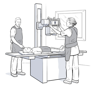
There may be a table in the room, and there will be a large X-ray camera suspended from the ceiling which can be moved in many directions to obtain various views. Portable X-ray equipment can be used to make films on patients in areas of the hospital, such as the operating room, the emergency department, or the intensive care unit.
A chest X-ray may be done in a standing, sitting, or lying position, depending on the condition of the child and the reason for the X-ray. For a standing or sitting film, your child will stand or sit in front of an X-ray plate. If the X-ray is taken in the lying position, the plate is placed beneath your child while he and she is lying on his and her back or side. If your child is an infant, he or she may be placed in a special device that will hold your child still while the X-ray is being taken.
The technologist will position your child properly in front of the plate, and then will step away to the controls of the machine. If your child is old enough to cooperate, he and she will be asked to take in a deep breath and hold it for a few seconds while the X-ray exposure is made. Otherwise, the technician will try to take the picture at the appropriate time by watching your child breathe.
In some situations, the doctor may want a film made from a side angle. This procedure is the same as the one just described, except that your child will stand, sit, or lie at a right or left angle to the X-ray plate and his or her arms will be raised out of the way.
Parents are usually able to stay in the room with their children to provide support and encouragement. You will be asked to wear a lead apron to protect you from unneeded exposure to radiation during the X-ray.
Portable X-ray machines may be used when it is difficult or unsafe to transport the child to the radiology department. Portable X-ray machines are generally used when the child is in an intensive care unit (ICU).
Depending on the results of the chest X-ray, additional tests or procedures may be scheduled to gather further diagnostic information.
Updated:
March 01, 2018
Sources:
A Manual of Laboratory and Diagnostic Tests. Fischbach, F. 2009, ed. 8, pp.764-5.
Reviewed By:
Bass, Pat F. III, MD, MPH,Grossman, Neil, MD,Image reviewed by StayWell medical illustration team.