Anatomy of the Skull Base
Anatomy of the Skull Base
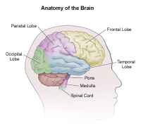
Your ability to run, jump, write with a pen, laugh, and experience pain all start in the brain, a mass of soft tissues and nerve cells attached to the spinal cord that sends messages throughout the body to let you move and feel. The brain is divided into several parts, all protected by the skull.
At the base of the skull is bone that supports 4 brain components—the frontal lobe, temporal lobe, brain stem, and cerebellum.
The skull base offers support from the bottom of the brain. Think of it as the floor of the skull, where the brain sits. Five bones make up the skull base. From front to back they are:
Frontal
Ethmoid
Sphenoid
Temporal
Occipital
Skull base tumors
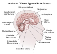
Tumors can form at the base of the skull or extend to the base of the skull after starting in another area of the body. This is called metastasis. The location of skull base tumors are often close to critical areas of the brain. This can make surgery difficult and potentially dangerous.
Skull base tumors may form in many areas, including the:
Meninges, the outer covering of the brain
Sinuses
Pituitary gland
Skull bone itself (osteosarcoma)
Symptoms of skull base tumors
Symptoms will vary, depending on the origin and site of the tumor. All symptoms tend to start slowly and progress gradually over time.
Tumors growing from the base of the cranium into the nose can cause symptoms similar to that of a chronic sinus infection:
Runny nose
Stuffy nose
Nosebleeds
Trouble breathing through the nose
Pressure in the face
Other types of skull base tumors may cause symptoms:
Blurry or double vision
Vision loss
Numbness in the top teeth
Bulging eyes
Tearing
Loss of smell
Loss of hearing
Headaches
Seizures
Nausea
Vomiting
Changes in mental status
Pain in the ear
Diagnosis
Skull base tumors can be diagnosed through:
Physical exam
Imaging tests, like MRI, PET, and CT scans
Biopsy
Treatment
Skull base tumors are hard to treat because of their location deep inside the brain. Treatment typically includes surgery when possible, followed by radiation therapy. Chemotherapy is sometimes used as a treatment option.
New surgical methods are currently being perfected to reach and remove skull base tumors that have been nearly unreachable through conventional surgery. One method, the endoscopic endonasal approach, allows surgeons to remove tumors through the nose. With another method, known as endoport surgery, a surgeon operates and removes a tumor through a strawlike tube inserted in a tiny hole drilled in the skull. It is then threaded into deep regions of the brain previously difficult, if not impossible, to reach. These and other minimally invasive procedures have led to better success rates in treating skull base cancers, with fewer complications and surgical side effects.
Key points to remember
Skull base tumors are very rare. But they can be dangerous and life threatening depending on the type, location, and size of the specific tumor. Advances in medical technology are making safe removal of these tumors more possible than ever before and are improving the outlook of people diagnosed with skull base tumors.
Updated:
January 10, 2018
Sources:
Patel CR., Skull Base Anatomy, Otolarynologic Clinics of North America (2016), 49(1); 9-20., Skull Base Anatomy, Elsevier, Inc., Uncommon Brain Tumors, Up To Date
Reviewed By:
Jasmin, Luc, MD,Watson, L. Renee, MSN, RN

