Bile Duct Cancer: Tests After Diagnosis
Bile Duct Cancer: Tests After Diagnosis
What tests might I have after being diagnosed?
After a diagnosis of bile duct cancer, you will likely need other tests. These tests help your healthcare providers learn more about the cancer. They can help show if the cancer has grown into nearby areas or spread to other parts of the body. The test results help your healthcare providers decide the best ways to treat the cancer. If you have any questions about these or other tests, be sure to talk with your healthcare team.
The tests you may have can include:
Endoscopic ultrasound
CT scan
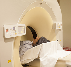
MRI
Cholangiography
Positron emission tomography (PET) scan
Imaging tests
Endoscopic ultrasound
Ultrasound tests use sound waves to create images of the inside of your body. For an endoscopic ultrasound, the doctor uses a thin, lighted tube called an endoscope. The endoscope is put in through your mouth and moved down into your intestine near the bile duct area. This lets the doctor get closer to the bile ducts to use ultrasound, which allows for more detailed images. Your doctor can use this test to see more clearly if the cancer has spread to nearby tissues.
CT scan
A CT scan can help your doctor see if bile duct cancer has spread to nearby lymph nodes or to other organs. A CT scan gives a better picture than an ultrasound. For the test, you lie still on a table as it slowly slides through the center of the ring-shaped CT scanner. The scanner sends a beam of X-rays at your belly (abdomen). A computer uses the data from the X-rays to create many detailed pictures of your insides. These are put together to create a 3-D picture. A CT scan doesn't hurt. You may be asked to hold your breath one or more times during the scan. In some cases, you may be asked to drink a contrast dye before the scan. You may be told to not to eat anything after drinking the contrast dye and before having the scan. The contrast dye will slowly pass through your system and come out in your bowel movements.
MRI
An MRI uses magnets, radio waves, and a computer to make detailed images of tissues and organs inside your body. MRIs may also be used to help see if cancer has spread outside of your bile ducts. Like a CT scan, an MRI can show more detail than X-rays. MRI is not necessarily better than a CT scan for bile duct cancer detection. But a new form of this test called magnetic resonance cholangiopancreatography (MRCP) gives clear and detailed images of this area in the body. Another test called MR angiography (MRA) is an MRI study of the blood vessels. It gives the best pictures of the blood vessels in the area. MRI may also be used instead of a CT scan for people who are allergic to contrast dye.
For this test, you lie still on a narrow table as it passes into a large steel tube that will scan your body. While inside the tube, radio waves are sent through your body to your abdomen. These are not X-rays. They are painless. A computer uses the data from the radio waves to create detailed pictures of the inside of your body. You may need more than one set of images. Each set may take 2 to 15 minutes. The whole scan may take an hour or more. There are loud, grating, and thumping noises during the scan. You may be given earplugs, headphones, or both to wear. If you are claustrophobic, you may be given a sedative before having this test. A 2-way intercom will let you talk to the people controlling the scanner at all times.
Cholangiography
A cholangiogram is a test that lets the doctor look at the bile ducts to see if they are narrow or blocked. There are different ways to do a cholangiogram. For example, magnetic resonance cholangiopancreatography (MRCP) creates detailed pictures of the bile ducts using the same type of machine used for an MRI. Other types of cholangiography include endoscopic retrograde cholangiopancreatography (ERCP) and percutaneous transhepatic cholangiography (PTC). These are more invasive. These also require the injection of a dye so the bile ducts can be seen on X-rays.
Positron emission tomography (PET) scan
For this test, a small amount of radioactive glucose (sugar) is injected into a vein. A scanner then creates pictures of the inside of the body and shows where the glucose is absorbed. These spots are called “hot spots.” Cancer cells absorb more glucose than normal cells, so this scan is used to show where cancer may have spread in the body. This test can also be used to show the difference between live cancer cells and scar tissue. A PET scan may be combined with a CT scan to provide more information. This is called a PET-CT scan.
Working with your healthcare provider
Your healthcare provider will talk with you about which tests you’ll have. Make sure to get ready for the tests as instructed. Ask questions and talk about any concerns you have.
Updated:
May 05, 2018
Sources:
NCCN Clinical Practice Guidelines in Oncology: Hepatobiliary Cancers Ver 3.2017 -- August 15, 2017. National Comprehensive Cancer Network.
Reviewed By:
LoCicero, Richard, MD,Stump-Sutliff, Kim, RN, MSN, AOCNS