Iridology
Iridology
Natural Standard Bottom Line Monograph, Copyright © 2013 (www.naturalstandard.com). Commercial distribution prohibited. This monograph is intended for informational purposes only, and should not be interpreted as specific medical advice. You should consult with a qualified healthcare provider before making decisions about therapies and/or health conditions.
While some complementary and alternative techniques have been studied scientifically, high-quality data regarding safety, effectiveness, and mechanism of action are limited or controversial for most therapies. Whenever possible, it is recommended that practitioners be licensed by a recognized professional organization that adheres to clearly published standards. In addition, before starting a new technique or engaging a practitioner, it is recommended that patients speak with their primary healthcare provider(s). Potential benefits, risks (including financial costs), and alternatives should be carefully considered. The below monograph is designed to provide historical background and an overview of clinically-oriented research, and neither advocates for or against the use of a particular therapy.
Related Terms
Angle of Fuch's, applied iridology, central heterochromia, cholesterol ring, ciliary zone, collarette, connective tissue type, Deck's pancreatic signs, density, flower, healing lines, inner-directed personality, iris biometrics, iris diagnosis, jewel, lacuna, left-brain development, lesion, lipemic diathesis, lipid ring, lymphatic rosary, nutritive zone, outer-directed personality, papillary margin, parasite lines, pigment ruff, pinguecula, prolapsus of transverse colon, psora, pterygium, pupil size, pupil sphincter muscle, radial furrows, radial solaris, radials, rarefaction, Rayid model, right-brain development, ring of determination, ring of harmony, ring of purpose, rings of freedom, sclerology, scurf ring, shading, shakers, sodium ring, stomach ring, stream, stress rings, tobacco snuffing, topo labile, topo stabile, trabeculae, transversal, vascular transversal, weak iris.
Background
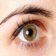
Iridology is the study of the iris (colored part of the eye) with the intention of gaining information about underlying diseases. Naturopaths and other practitioners may use this technique.
Iridology is based on the belief that each body region and organ is represented at one specific location in the iris. Abnormalities in a region are suggested to correspond to abnormalities in the respective organ. This technique was developed in Budapest, Hungary in the 1800s by the physician Ignatz von Pezcely. Iridology was later adapted by Bernard Jensen, an American chiropractor. Jensen based his practice on detailed diagrams of the left and right irises, assigning every organ, many body parts, and several bodily functions to a specific location on one or both irises.
The popularity of iridology grew in the 1900s. Iridologists visually access the iris of their patients by direct examination or by studying close-up photographs. Some iridologists use sclerology, a technique that studies lines on the sclera (the white part of the eye), which they believe can show changes in health patterns and conditions. The Rayid model may also be used, which studies eye patterns with the aim to evaluate mental, emotional, spiritual, and physical balance.
Theory
Iridologists believe that the degrees of light and darkness in the iris give clues to the body's general health. An iridologist may use a special camera to take pictures of a client's iris or may examine it using an ophthalmoscope. Some iridologists take color photographs or transparencies, which they magnify and read. Microscopes or computer imaging may be used.
By studying these images, practitioners try to identify weaknesses in the body or diagnose disease. Health status is analyzed by studying colors, marks, and signs in the iris, the pupil, and the sclerae of the eye. Pigmentation changes and potential drug deposits are studied. It is believed that diseases may show up as pigment changes, light or dark streaks, flecks, or spots. Textures of "fibers" in the iris are also studied. According to the International Iridology Research Association, iridology cannot name a disease; only a medical doctor can.
Three basic types of diagnosis categories may be used by iridologists to narrow down disease states of patients. (1) Lymphatic (blue or blue grey): Iridologists believe that these patients may be prone to immune system problems, allergies, asthma, ezcema, upper respiratory tract infections, tonsillitis, and arthritis. The status of the lungs, mucous membranes, urogenital tract, kidneys, joints, and adrenal glands are believed to be seen here. (2) Hematogenous (brown): Iridologists believe that these patients may have lowered blood cell counts, anemia, glandular, and circulatory disorders. (3) Biliary (blue and brown): Iridologists believe that these patients may have allergies, disorders of the liver, gallbladder, or digestive system, and abnormalities with blood sugar or calcium metabolism. The circulatory system, spleen, bone marrow, and endocrine glands are believed to be reflected here.
Different colors seen within the iris are believed to represent different aspects of health. For example, yellow may be interpreted as reflecting poor kidney function. Rings in the eye are also believed to play a role in diagnosis.
Conditions diagnosed via these methods may be treated by iridologists, depending on their type of training, or they may recommend another therapist. Iridologists may also counsel clients about nutrition and lifestyle.
Scientific Evidence
Uses These uses have been tested in humans or animals. Safety and effectiveness have not always been proven. Some of these conditions are potentially serious, and should be evaluated by a qualified healthcare provider. |
Grade* |
Hypertension (diagnosis) Preliminary studies suggest that iridology may assist in the identification of individual predispositions for vascular diseases such as hypertension (high blood pressure). Further research is needed to confirm these findings. |
C |
Cancer (diagnosis) There is currently limited available data supporting iridology as a diagnostic tool in cancer. Additional study is needed. |
D |
Gallbladder disease (diagnosis) Preliminary study examined the ability of iridologists to diagnose gallbladder disease from slide photographs of patients with the disease and found no evidence of agreement or diagnostic accuracy. There is no evidence supporting iridology as a diagnostic tool in gallbladder disease. |
D |
Kidney disease (diagnosis) Preliminary study submitted photographs of irises of kidney disease patients to practicing iridologists and found no evidence of accurate detection of kidney disease. There is no evidence supporting iridology as a diagnostic tool in kidney disease. |
D |
*Key to grades:A: Strong scientific evidence for this use; B: Good scientific evidence for this use; C: Unclear scientific evidence for this use; D: Fair scientific evidence against this use (it may not work); F: Strong scientific evidence against this use (it likely does not work). |
|
Tradition/Theory
The below uses are based on tradition or scientific theories. They often have not been thoroughly tested in humans, and safety and effectiveness have not always been proven. Some of these conditions are potentially serious and should be evaluated by a qualified health care professional.
Diagnosis of: allergies, anemia, arthritis, asthma, circulatory disorders, cough, diabetes, digestive system disorders, eczema, endocrine disorders, glandular disorders, heart disease, immune system problems, intestinal disorders, liver disease, low white blood cell counts, lung disease, problems with blood sugar metabolism, psoriasis, stomach disorders, tonsillitis, ulcerative colitis, under-active pancreas, upper respiratory tract infections, urinary stones.
Safety
Many complementary techniques are practiced by healthcare professionals with formal training, in accordance with the standards of national organizations. However, this is not universally the case, and adverse effects are possible. Due to limited research, in some cases only limited safety information is available.
Iridology is generally a non-invasive technique. However, it may pose a danger if it is used for disease diagnosis instead of more proven approaches. Incorrect diagnoses have been reported using this method and can potentially lead to inappropriate treatment or to psychological stress in patients. In addition, serious medical problems may go undiagnosed. Iridology is therefore not recommended as a sole method of diagnosis or treatment for any condition.
Author Information
This information is based on a systematic review of scientific literature edited and peer-reviewed by contributors to the Natural Standard Research Collaboration (www.naturalstandard.com).
References
Natural Standard developed the above evidence-based information based on a thorough systematic review of the available scientific articles. For comprehensive information about alternative and complementary therapies on the professional level, go to www.naturalstandard.com. Selected references are listed below.
Bartholomew RE, Likely M. Subsidising Australian pseudoscience: is iridology complementary medicine or witch doctoring? Aust N Z J Public Health 1998;22(1):163-164.
Berggren L. Iridology. A critical review. Acta Ophthalmol 1985;63(1):1-8.
Buchanan TJ, Sutherland CJ, Strettle RJ, et al. An investigation of the relationship between anatomical features in the iris and systemic disease with reference to iridology. Complement Ther Med 1996;4:98-102.
Caradonna B. Western medicine looks at iridology again. Iridol Rev J 1990;2(2):13-14.
Caradonna B. Iridology research part 1-design. Iridol Rev J 1994;2(3):3-5.
Cockburn D. A study of the validity of iris diagnosis. Austral J Optom 1981;64(4):154-157.
Davidson F. How iridology and orthodox medicine work together. J Altern Med 1985;3(11):17-18.
Ernst E. Iridology: not useful and potentially harmful. Arch Ophthalmol 2000;118(1):120-121.
Friedman GD, Selby JV, Quesenberry CP, et al. Eye color and hypertension. Med Hypoth 1990;33(3):201-206.
Kleinstein RN, Seitz MR, Barton TE, et al. Iris color and hearing loss. Am J Optom Physiol Opt 1984;61(3):145-149.
Knipschild P. Looking for gall bladder disease in the patient's iris. BMJ 1988;297(6663):1578-1581. View Abstract
Popescu MP, Waniek DA. [Improved irido-diagnostic method: possibilities of computerized iridology]. Rev Chir Oncol Radiol 1986;30(1):29-33.
Simon A, Worthen DM, Mitas JA. An evaluation of iridology. JAMA 1979;242(13):1385-1389. View Abstract
Worrall RS. Pseudoscience--a critical look at iridology. J Am Optom Assoc 1984;55(10):735-739. View Abstract
Yoo CS, Hwang WJ, Hong SH, et al. Relationship between iris constitution analysis and TNF-alpha gene polymorphism in hypertensives. Am J Chin Med 2007;35(4):621-629. View Abstract
Copyright © 2013 Natural Standard (www.naturalstandard.com)
The information in this monograph is intended for informational purposes only, and is meant to help users better understand health concerns. Information is based on review of scientific research data, historical practice patterns, and clinical experience. This information should not be interpreted as specific medical advice. Users should consult with a qualified healthcare provider for specific questions regarding therapies, diagnosis and/or health conditions, prior to making therapeutic decisions.
Updated:
March 22, 2017