Focal dermal hypoplasia
Focal dermal hypoplasia
Natural Standard Monograph, Copyright © 2013 (www.naturalstandard.com). Commercial distribution prohibited. This monograph is intended for informational purposes only, and should not be interpreted as specific medical advice. You should consult with a qualified healthcare provider before making decisions about therapies and/or health conditions.
Related Terms
Anhidrosis, combined mesoectodermal dysplasia, DHOF, ectodermal and mesodermal dysplasia congenital, ectodermal and mesodermal dysplasia with osseous involvement, FDH, focal dermal dysplasia syndrome, focal dermato-phalangeal dysplasia, FODH, Goltz syndrome, Goltz-Gorlin syndrome, PORCN gene.
Background
Focal dermal hypoplasia (FDH) is an ectodermal dysplasia, one of a group of disorders that affect the outer layer of the developing embryo. This layer, called the embryonic ectoderm, develops into many parts of a baby's body, including the eyes, skin, teeth, and bones. In ectodermal dysplasias, these parts do not develop normally.
The main symptoms associated with FDH are skin problems, which may include streaks or lines of tumorlike growths on various parts of the body. These are usually present at birth but may develop over time. Older patients with FDH may have pain associated with skin problems. Other symptoms may affect the urinary, digestive, cardiovascular, and central nervous systems.
FDH is caused by a mutation or defect in the PORCN gene. While the exact function of this gene is unknown, activity of the PORCN gene has been detected in the long bones, facial bones, fingers, toes, and teeth. About 90% of cases of FDH are inherited or passed down among family members. FDH is inherited as an X-linked dominant trait.
FDH is a rare condition, with only about 200 to 300 known cases worldwide. The exact incidence of the disorder is unknown. No racial or ethnic group is affected more than any other.
About 90% of people with FDH are female. Males with FDH generally die before birth. Individuals who are severely affected by FDH may die in infancy. Those who are mildly affected may have a normal life expectancy.
Risk Factors
Because focal dermal hypoplasia (FDH) is inherited, the only known risk factor is a family history of the disease. FDH is a rare condition, with only about 200 to 300 known cases worldwide. The exact incidence of the disorder is unknown. No racial or ethnic group is affected more than any other.
About 90% of people with FDH are female. Males with FDH generally die before birth. Individuals who are severely affected by FDH may die in infancy. Those who are mildly affected may have a normal life expectancy.
Causes
Genetic mutations: Focal dermal hypoplasia (FDH) is caused by a mutation or defect in the PORCN gene. While the exact function of this gene is unknown, activity of the PORCN gene has been detected in the long bones, facial bones, fingers, toes, and teeth.
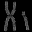
X-linked dominant inheritance: About 90% of cases of FDH are inherited or passed down among family members. FDH follows an X-linked dominant pattern of inheritance, meaning that the defective PORCN gene is located on the X chromosome and that only one copy of the defective gene is required for the disease to appear.
Females have two copies of the X chromosome, but males have one X chromosome and one Y chromosome. Males inherit an X chromosome from the mother and a Y chromosome from the father, so a male can inherit the defective gene only from the mother. About 90% of people with FDH are female. Males with FDH generally die before birth.
Random occurrence: FDH may occur in individuals with no family history of the disorder. This is the result of a spontaneous mutation in the sperm or egg cells or in the developing embryo.
Signs and Symptoms
General: Symptoms of focal dermal hypoplasia (FDH) are generally present at birth. Most symptoms do not worsen with age, although skin-related symptoms may change over time.
Bone: People with FDH may have skeletal abnormalities, including problems with the fingers and toes. For example, fingers and toes may be fused or abnormally curved sideways, or there may be missing or extra fingers and toes. Other patients may have claw-like hands and feet that are split down the middle. The bones also tend to be striated, or lined.
Additional symptoms may include short stature, sloping shoulders, underdeveloped bones in the pelvis, scoliosis (abnormal sideways curvature of the spine), or kyphosis (forward curvature of the spine). Osteopenia, or low bone density, may also be present. Giant cell-like tumors have been seen in the bones in FDH. However, none have been reported as malignant.
Central nervous system: The central nervous system (CNS), which consists of the brain and spinal cord, may also be affected in FDH. Problems may include a breakdown of part of the brain, a small head, cysts in the brain or the lining surrounding the brain, or collection of fluid in the brain.
Cognitive: Some people with FDH may have intellectual disabilities.
Dental: People with FDH may have fingerlike tumors on the lips. In addition, they may have underdeveloped, abnormally small, or missing teeth. There may also be problems with the alignment of the jaw and teeth. In rare cases, patients may have a cleft palate, or incomplete closure of the roof of the mouth.
Eyes: Small holes (colobomas) or cysts (hydrocystomas) may be present on parts of the eye. In addition, patients may have strabismus (crossed eyes), abnormally small eyes, and nystagmus (involuntary eye movements).
Face: The face may appear asymmetrical. Ears are often protruding and low-set and may be of different sizes. People with FDH may also have a narrow bridge and wide tip of the nose. A pointed chin is also common in FDH.
Hair: As with most ectodermal dysplasias, the hair, eyebrows, and eyelashes tend to be sparse and brittle. Some people may experience hair loss or patchy or compete baldness.
Hearing: People with FDH may have impaired hearing, which may be caused by structural problems with the skull or cholesteatoma (a growth in the ear canal).
Nails: The nails may be poorly shaped or underdeveloped, have streaks or grooves, or be spoon-shaped.
Skin: The most common feature of FDH is streaks or lines of tumor-like growths on various parts of the body, especially the thighs, forearms, and cheeks. These may be painless or tender to the touch. In addition, there may be collections of fat cells, which give a lumpy or pouchy appearance to the skin. Individuals may also have papillomas (small fingerlike tumors) on the mucous membranes, such as the inside of the mouth, nose, and anus, as well as the lips, fingers, toes, and groin. Skin may also have too much or too little pigment in some areas. Some patients with FDH may develop telangiectases, which are collections of expanded blood vessels under the skin. Other symptoms of FDH may include anhidrosis (a reduced or absent ability to sweat).
Other: In rare cases, people with FDH may have problems with the heart, intestines, and urinary system, and may be prone to infection.
Diagnosis
General: Focal dermal hypoplasia (FDH) may be diagnosed following a thorough family history and complete physical exam. FDH may be suspected based on observation of the distinctive physical characteristics associated with the condition, specifically, dead areas of skin; fat nodules in the skin, which resemble soft, yellow-pink bumps; and pigment changes. The nails can be ridged, and hair can be sparse or absent. Limb malformations include fusion of some of the fingers. Craniofacial findings can include facial asymmetry, cleft lip and palate, and pointed chin.
Biopsy: In a biopsy, a very small sample of tissue is taken for examination. For diagnosis of FDH, a biopsy of the skin may be taken to assess the type and severity of skin conditions and to look for the presence of sweat glands, fat deposits, collections of blood vessels, the absence or excess of pigment, and the different types of tumors that may develop in FDH.
Imaging: Imaging studies such as X-rays may be used to assess the skeleton in people with FDH. These can help identify striation (lines) on the bones. Although these are not specific to FDH, their detection may help on making a diagnosis. Ultrasound may be used in pregnant women with a family history of FDH. Ultrasound may identify growth delays, problems with specific organs, and developmental problems.
Genetic testing: If FDH is suspected, a cytogenetic test may be performed to confirm a diagnosis. A sample of the patient's blood is taken and analyzed in a laboratory for the defect in the PORCN gene. If this defect is detected, a positive diagnosis is made.
Prenatal DNA testing: If there is a family history of FDH, prenatal testing may be performed to determine whether the fetus has the disorder. Amniocentesis and chorionic villus sampling (CVS) can diagnose FDH. However, because there are serious risks associated with these tests, patients should discuss the potential health benefits and risks with a medical professional.
During amniocentesis, a long, thin needle is inserted through the abdominal wall and into the uterus, and a small amount of amniotic fluid is removed from the sac surrounding the fetus. Cells in the fluid are then analyzed for normal and abnormal chromosomes. This test is performed after 15 weeks of pregnancy. The risk of miscarriage is about one in 200-400 patients. Some patients may experience minor complications, such as cramping, leaking fluid, or irritation where the needle was inserted.
During chorionic villus sampling (CVS), a small piece of tissue (chorionic villi) is removed from the placenta between the ninth and 14th weeks of pregnancy. CVS may be performed through the cervix or through the abdomen. The cells in the tissue sample are then analyzed for the mutation in the PORCN gene. Miscarriage occurs in about 0.5%-1% of women who undergo this procedure. Prenatal testing is possible for women with high-risk pregnancies if the disease-causing mutation in the family has been identified.
Complications
Complications of focal dermal hypoplasia (FDH) tend to be mild and associated with functional issues. Large benign tumors near the mouth or anus may physically interfere with eating and elimination. In rare cases, tumors in the digestive tract may block the passage of food or waste products. In some cases, these tumors are mistaken for viral warts, such as those located near the anus or genitals, and improperly managed.
Skin lesions: FDH can cause painful erosive lesions that are prone to infection if improperly managed.
Hand and foot malformations: FDH causes hand and foot malformations, which may result in difficulty in performing everyday tasks, including walking.
Treatment
General: There is no known cure for focal dermal hypoplasia (FDH). Instead, treatment aims to reduce symptoms and prevent or treat complications. Patients with FDH should be regularly seen by a pediatrician, dermatologist, audiologist, ophthalmologist, and various surgeons, based on their individual symptoms.
Dental care: Patients with FDH must practice good preventive dental care, including regular flossing, teeth brushing, and visits to the dentist. Dentures may be appropriate for patients with FDH who are missing teeth.
Education: By law, patients with FDH syndrome who suffer from intellectual disabilities must have access to education that is tailored to their specific strengths and weaknesses. According to the Individuals with Disabilities Education Act, all children with disabilities must receive free and appropriate education. This law states that staff members of the patient's school must consult with the patient's parents or caregivers to design and write an individualized education plan based on the child's needs. The school faculty must document the child's progress in order to ensure that the child's needs are being met.
Educational programs vary among patients, depending on the child's specific learning disabilities. In general, most experts believe that children with disabilities should be educated alongside their nondisabled peers. The idea is that nondisabled students will help the patient learn appropriate behavioral, social, and language skills. Therefore, some FDH patients are educated in mainstream classrooms. Others attend public schools but take special education classes. Still others attend specialized schools that are equipped to teach children with disabilities.
Laser therapy: Treatment with a laser may improve the appearance of the skin.
Physical therapy: FDH causes hand and foot malformations, which may result in difficulty in performing everyday tasks, including walking. Physical therapy can help manage these symptoms.
Speech language therapy: Some patients with FDH may benefit from speech-language therapy, because these individuals often develop communication skills more slowly than normal. During speech-language therapy, a qualified speech-language professional (SLP) works with the patient on a one-to-one basis, in a small group, or in a classroom, to help the patient improve speech, language, and communication skills. Programs are tailored to the patient's individual needs.
Speech pathologists use a variety of exercises to improve the patient's communication skills. Exercises typically start simple and become more complex as therapy continues. For instance, the therapist may ask the patient to name objects, tell stories, or explain the purpose of an object.
On average, patients receive five or more hours of therapy per week for three months to several years. Doctors typically recommend that treatment be started early to ensure the best possible prognosis for the child.
Surgery: Surgery may be necessary if skeletal or oral problems are severe. Surgical management of papillomas, the fingerlike tumors that occur in and around the mouth, anus, and genitals, is not permanent and may need to be repeated, as new ones may form.
Integrative Therapies
Currently there is a lack of scientific evidence on the use of integrative therapies for the treatment or prevention of focal dermal hypoplasia (FDH).
Prevention
General: Because focal dermal hypoplasia (FDH) syndrome is an inherited condition, there is currently no known way to prevent the disease.
Genetic testing and counseling: Individuals who have FDH may meet with a genetic counselor to discuss the risks of having children with the disease. Genetic counselors can explain the options and the associated risks of various tests, including preimplantation genetic diagnosis (PGD), amniocentesis, and chorionic villus sampling (CVS).
Preimplantation genetic diagnosis (PGD) may be used with in vitro (artificial) fertilization. In PGD, embryos are tested for the defective PORCN gene, and only the embryos that are not affected may be selected for implantation. Because FDH can be detected in a fetus, parents may choose whether to continue the pregnancy. Genetic counselors may assist parents with these difficult decisions.
Author Information
This information has been edited and peer-reviewed by contributors to the Natural Standard Research Collaboration (www.naturalstandard.com).
Bibliography
Natural Standard developed the above evidence-based information based on a thorough systematic review of the available scientific articles. For comprehensive information about alternative and complementary therapies on the professional level, go to www.naturalstandard.com. Selected references are listed below.
Bellosta M, Trespiolli D, Ghiselli E, et al. Focal dermal hypoplasia: report of a family with 7 affected women in 3 generations. Eur J Derm. 1996;6:499-500. View Abstract
Feinberg A, Menter MS. Focal dermal hypoplasia (Goltz syndrome) in a male: a case report. S Afr Med J. 1976;50:554-5. View Abstract
Goltz RW. Focal dermal hypoplasia syndrome: an update. Arch Derm. 1992;128:1108-11. View Abstract
Gordjani N, Herdeg S, Ross UH, et al. Focal dermal hypoplasia (Goltz-Gorlin syndrome) associated with obstructive papillomatosis of the larynx and hypopharynx. Eur J Derm. 1999;9:618-20. View Abstract
Larreque M, Duterque M. Striated osteopathy in focal dermal hypoplasia. Arch Derm. 1975;111:1365. View Abstract
National Foundation for Ectodermal Dysplasias. www.nfed.org.
Natural Standard: The Authority on Integrative Medicine. www.naturalstandard.com.
Wang X, Sutton VR, Peraza-Llanes JO, et al. Mutations in X-linked PORCN, a putative regulator of WNT signaling, cause focal dermal hypoplasia. Nat Genet. 2007;39:836-8. View Abstract
Copyright © 2013 Natural Standard (www.naturalstandard.com)
The information in this monograph is intended for informational purposes only, and is meant to help users better understand health concerns. Information is based on review of scientific research data, historical practice patterns, and clinical experience. This information should not be interpreted as specific medical advice. Users should consult with a qualified healthcare provider for specific questions regarding therapies, diagnosis and/or health conditions, prior to making therapeutic decisions.
Updated:
March 22, 2017
