Understanding Endoscopic Endonasal Surgery
Understanding Endoscopic Endonasal Surgery
Endoscopic endonasal surgery is a type of procedure to treat problems, such as a tumor, in the front or bottom of the brain or top of the spinal cord. A thin tube called an endoscope is used to let the surgeon do the surgery. Instead of cutting through the skull, a surgeon can put the scope and tiny tools through the nose. This lets the healthcare provider reach areas that are hard to reach in other ways. Recovery is also faster and less painful than with open surgery.
How to say it
EN-doh-skah-pik EN-doh-nayz-uhl
Why endoscopic endonasal surgery is done
This kind of surgery may be done to:
-
Remove a tumor or other growth in the pituitary gland, sinuses, or brain
-
Stop leakage of cerebrospinal fluid (CSF)
-
Treat an infection that won’t go away
-
Treat problems affecting the optic nerve
-
Repair a problem with blood vessels in the brain called arteriovenous malformation (AVM)
How endoscopic endonasal surgery is done
The treatment is done in a hospital, and may take several hours. During the procedure:
-
You lie on a surgery table. You’re given medicine to make you sleep through the treatment.
-
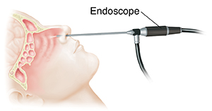
The surgeon puts a thin, stiff tube called an endoscope into your nose and sinuses. The scope is narrow and fits through natural gaps in these areas. The scope has a light and a camera. Images from the camera are sent to a computer screen.
-
The surgeon also puts small surgery tools through your nose. This is often done through the same nostril as the scope. The tools are used to remove a tumor or treat the problem area as needed. The surgeon may remove or cut parts of bone on the underside of the skull or other tissue during the treatment.
-
When the surgery is done, the surgeon removes the tools and scope. Your nose and sinuses may be filled with nasal packing. This is material that will be removed in a day or two. You’ll be in the hospital one or more nights after surgery.
-
A newer technique offers 3-D viewing to give the surgeon a better understanding of the space and what he or she is seeing on a computer screen.
Risks of endoscopic endonasal surgery
The risks of endoscopic endonasal surgery include:
-
CSF leaking from the nose
-
Excess bleeding
-
Damage to blood vessels and nerves in the area
-
Pain
-
Crusting of the inside of the nose and sinuses that lasts for weeks or months
-
Trouble breathing through the nose
-
Reaction to anesthesia
-
Infection of the covering of the brain and spinal cord (meninges)
Updated:
September 17, 2019
Sources:
Chan J, et al. Endoscopic Endonasal Approaches to the Skull Base and Paranasal Sinuses. In: Brackmann DE, editor. Otologic Surgery. 4 ed. Philadelphia: Elsevier; 2016. p. 589-603., Jho DH, et al. Endoscopic Endonasal Pituitary and Skull Base Surgery. In: Quiñones-Hinojosa A, editor. Schmidek and Sweet's Operative Neurosurgical Techniques. 6 ed. Philadelphia: Elsevier; 2012. p. 257-79., Pasricha PJ, et al. Natural orifice transluminal endoscopic surgery (NOTES). Up To Date April 10 ed: Up To Date; 2015. p. 8., Verillaud B, et al. Endoscopic endonasal skull base surgery. European Annals of Otorhinolaryngology, Head and Neck Diseases. 2012 August 1;129(4):190-6.
Reviewed By:
Ashutosh Kacker MD,Anne Fetterman RN BSN,Raymond Kent Turley BSN MSN RN