The Day of Your Craniotomy
The Day of Your Craniotomy
Craniotomy is a surgical opening made in the skull for treatment of several types of problems in the brain. Special tools are used to remove a piece of the skull and allow access to the brain for surgical treatment. The most commons reasons for having a craniotomy include a blood clot, tumors, aneurysms and arteriovenous malformations (AVM), and brain abscess.
Arrive at the hospital on time. You may still have concerns and are likely to feel a bit nervous. Your healthcare team will try to answer all your questions. They will also do all they can to put you at ease.
Just before surgery
The healthcare provider in charge of your anesthesia will talk with you before surgery. You will be given general anesthesia to help you “sleep” through the surgery. At some point, an IV (intravenous) line is placed in your arm. This line can supply medicine and fluids as needed. In many cases, some or all of your hair is shaved. This is done to decrease the risk of infection.
Reaching the brain
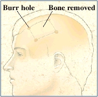
The surgeon makes an incision in your scalp. Then burr holes are drilled in the skull. The bone between the holes is cut and lifted away. The surgeon then opens the dura (meninges), exposing the brain. The next step will depend on your specific problem. In some cases, certain nerves may be stimulated while the response in the brain is monitored. This is done to make sure that normal brain tissue is not disturbed during the surgery.
Finishing the craniotomy
When the goal of surgery is met, the dura covering the brain is closed. Sometimes, a dura substitute or dural sealants ares used to close the meninges. Usually, the skull bone is put back and held in place with wire mesh,or screws and metal plates. If there is too much brain swelling, your surgeon might choose not to put back the bone flat. It can be put back at a later time once the swelling has gone down. On occasion, a metal mesh is used if the bone cannot be used again or if there is a bone infection (known as osteomyelitis). If blood or fluid remains in the brain tissue, a drain may be placed through a burr hole for a few days. Most of the time, the burr holes are filled or covered with metal plates right before closing the skin. Then the skin incision is closed with stitches or staples.
Risks of surgery
Risks of a craniotomy are:
Seizure (jerking movements or loss of consciousness)
Infection
Loss of memory or confusion
Swelling or bleeding in the brain
Blood clots
Loss of sensation, including vision
Weakness or paralysis
Death
Note to family and friends
You can wait in a nearby room during surgery. Craniotomy often takes 3 to 5 hours, or more, to complete. If possible, be sure to leave a phone number to receive any news, or have someone stay in the waiting room. The doctor will come talk with you as soon as surgery is over. You’ll also be told when you can visit your loved one.
Updated:
December 31, 2017
Reviewed By:
Jasmin, Luc, MD,Sather, Rita, RN
