Having Contrast Echocardiography
Having Contrast Echocardiography
Your healthcare provider recommends that you have contrast echocardiography (also called “a contrast echo”). This is an imaging test that uses sound waves (ultrasound) to take pictures of the heart while it is beating. During the test, a special dye (contrast agent) is injected into your vein to help show structures in the heart better. It allows the inside of the heart to be seen more clearly on the ultrasound pictures. A contrast echo is most often done to check for certain heart problems that can't be detected with other imaging tests. From start to finish, the test takes about 60 minutes. An echocardiogram lets your provider see how well the heart muscle is working, check the size of the heart chambers, and see how the valves are functioning.
Before the test
Follow any instructions given by your healthcare provider to prepare for the test.
You may be asked not to eat a heavy meal before the test to improve the images of the heart.
On the day of the test, wear a two-piece outfit. You may need to undress from the waist up and put on a short hospital gown.
Let the technologist know
For your safety, let the technologist know if you:
Have a known or suspected heart or lung problem
Have any other medical conditions
Are allergic to perflutren (contrast agent), blood, blood products, albumin, or any medicines
Are taking any medicines, including vitamins, herbs, and over-the-counter medicines
Are or may be pregnant
Are breastfeeding
During the test
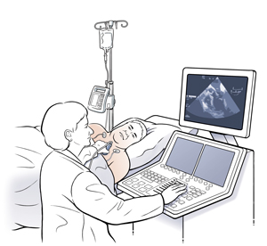
A contrast echo is done by an ultrasound technologist (sonographer). A nurse or doctor (cardiologist) may also be present during the test.
You’ll lie on your back on an exam table or hospital bed.
An IV line is placed into a vein in your arm or hand.
Small pads (electrodes) are placed on your chest to monitor your heart rate. In addition, special equipment is used to monitor your vital signs (blood pressure, breathing, and blood oxygen level).
A cool, clear or blue colored gel is placed on your chest. Then a handheld device called a transducer is moved firmly over your chest. The transducer releases harmless sound waves, which are processed by a computer. Live pictures of your heart can then be viewed on a monitor.
The contrast agent is injected through the IV into your bloodstream to reach your heart.
At times, you may be asked to change positions. This allows pictures of your heart to be taken from different angles. You may also be asked to hold your breath for a few seconds. This helps prevent the pictures from being blurry.
When the test is complete, the gel is cleaned off your chest and the IV is removed from your arm or hand.
The pictures of your heart are sent to your healthcare provider or a cardiologist for review.
After the test
You may need to rest after the test. Your vital signs will continue to be monitored during this time. You’ll be told when you can go home.
After you go home, return to your usual activities when you feel ready.
Risks and possible complications
Risks and possible complications of a contrast echo include:
Excessive bruising, bleeding, swelling, or other problems at the IV site
Tenderness on the chest wall from pressing the transducer against the tissue
Side effects such as dizziness, weakness, fatigue, palpitations, headaches, and nausea
Allergic reaction to the contrast (rash, hives, trouble breathing, swelling of the mouth, face, lips, or tongue). If you have an allergic reaction to the contrast, report it immediately and call 911 for any breathing difficulties.
Follow-up care
Follow up with your healthcare provider, or as advised. He or she will go over the test results with you during a follow-up visit. This is usually within 1 to 2 weeks. Be sure to share any concerns you have with your provider.
Updated:
September 02, 2017
Sources:
Appis, A., Update on the safety and efficacy of commercial ultrasound contrast agents in cardiac applications, Echo Research and Practice (2015); R55-R62, Contrast echocardiography: Contrast agents, safety, and imaging technique, Up To Date, Lepper, W., Myocardial Contrast Echocardiography, circulation (2004); 109; 3132-3135, Mulvagn, SL. American Society of Echocardiography Consensus Statement on the Clinical Applications of Ultrasonic Contrast Agents in Echocardiography. Journal of the American Society of Echocardiography (2008); 21(11); pp. s1179-s12-1
Reviewed By:
Gandelman, Glenn, MD, MPH,Image reviewed by StayWell medical illustration team.,Snyder, Mandy, APRN