Macular degeneration
Macular degeneration
Natural Standard Monograph, Copyright © 2013 (www.naturalstandard.com). Commercial distribution prohibited. This monograph is intended for informational purposes only, and should not be interpreted as specific medical advice. You should consult with a qualified healthcare provider before making decisions about therapies and/or health conditions.
Related Terms
Age-related macular degeneration, AMD, Amsler grid, angiography, antioxidants, atrophic, autofluorescence, autosomal dominant hemorrhagic macular dystrophy, Best's vitelliform macular dystrophy, CFH, Charles Bonnet syndrome, choroidal neovascularization, CNV, compliment factor H, corticosteroid, disciform degeneration, Doyne's honeycomb retinal dystrophy, drusen, drusenoid, fundus flavimaculatus, implantable optical device, JMD, juvenile macular degeneration, macula, malattia levintinese, mydriasis, mydriatic, nonexudative, obesity, OCT, ophthalmologist, optic nerve, optical coherence tomography, photocoagulation, photodynamic therapy, retina, retinal pigment epithelial detachment, retinal pigment epithelium, rheophoresis, RPE, smoking, Sorsby's fundus dystrophy, Stargardt macular dystrophy, subretinal neovascularization, ultraviolet, UV, vascular endothelial growth factor, VEGF, Yannuzzi card.
Background
Macular degeneration is a degenerative disease of the retina (a thin layer of nerve cells that lines the back of the eyeball) that causes progressive loss of central vision.
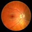
Central vision loss is due to the deterioration of the central part of the retina, known as the macula. The macula is involved in detailed vision. Light sensing cells in the macula, known as photoreceptors, convert light into electrical impulses. Then the impulses are transferred to the brain via the optic nerve. Central vision loss from macular degeneration occurs when photoreceptor cells in the macula degenerate.
The risk of developing macular degeneration increases with age. The disease most often affects people in their sixties and seventies. Macular degeneration is the most common cause of vision loss in individuals over the age of 60.
Individuals with macular degeneration may first notice a blurring of central vision that is most apparent when performing visually detailed tasks such as reading and sewing. Blurred central vision may also make straight lines appear slightly distorted or warped. As the disease progresses, blind spots form within central vision. In most cases, if one eye has macular degeneration, the other eye will also develop the disease. The extent of central vision loss varies according to the type of macular degeneration. Individuals can adapt to peripheral vision, or vision out of the corners of the eye, although some visual impairment will exist without central vision.
Age-related macular degeneration is the most common type of macular degeneration and is the leading cause of legal blindness in people older than 60 years in the United States. Most individuals with macular degeneration retain peripheral vision and learn to optimize the use of their remaining vision. Legally blind individuals are those whose best visual acuity or sharpness (with glasses or contact lenses, if needed) is 20/200 or worse in their better eye, or whose visual field, regardless of acuity, is restricted to a 20 degree diameter (10 degree radius).
The disease affects more than 10 million Americans, including 23% of Americans older than 90 years. Because overall life expectancy continues to increase, age-related macular degeneration has become a major public health problem.
Deficiencies in antioxidants (specifically zinc and vitamins A, C, and E) have been noted in some people with age-related macular degeneration. Antioxidants may protect against age-related macular degeneration by preventing free radicals or unstable oxygen from damaging the retina. Antioxidants can be found in foods such as green, leafy vegetables, vegetables of color (oranges, yellows, purples), and fruits. Dietary and lifestyle changes are necessary, including eating less fat (from meats and dairy products) and eating more vegetables and fruits, increasing exercise, and weight control.
There are several forms of macular degeneration that affect children, teenagers, or adults that are known as early onset or juvenile macular degeneration. Many of these forms are hereditary and are more accurately called macular dystrophies, instead of degenerations. Stargardt macular dystrophy is the most frequent type of juvenile-onset form of macular dystrophy. Other types include Best's vitelliform macular dystrophy, Doyne's honeycomb retinal dystrophy, Sorsby's fundus dystrophy, malattia levintinese, fundus flavimaculatus, and autosomal dominant hemorrhagic macular dystrophy.
Individuals affected with Stargardt's first present with the complaint of a decrease in central vision, generally within the first 20 years of life. In some instances, persons may not note visual impairment until their 30's or 40's. In the majority of instances, individuals do not experience a loss in peripheral vision or night-blindness, although it has been reported that persons with Stargardt's have some problems seeing in the dark.
Types of Age-Related Macular Degeneration
There are two types of age-related macular degeneration (AMD): dry and wet.
Dry form: Dry macular degeneration is sometimes called atrophic, nonexudative, or drusenoid macular degeneration. More than 90% of all people with AMD have the dry form. With dry macular degeneration, yellow-white deposits, called drusen, accumulate in the retinal pigment epithelium (RPE) tissue beneath the macula. Drusen deposits are composed of waste products from photoreceptor cells. For unknown reasons, RPE tissue can lose its ability to process waste, which is normally flushed out of the eye into lymphatic tissue. As a result, drusen deposits accumulate in the RPE. Drusen deposits are typically present in patients with dry macular degeneration. These deposits are thought to interfere with the function of photoreceptors in the macula, causing progressive degeneration of these cells. Drusen deposits can, however, be present in the retina without vision loss.
Vision loss from dry macular degeneration occurs very gradually over the course of many years (sometimes decades). Central vision, or straight ahead vision, in the background may remain stable between annual eye examinations. Individuals with macular degeneration do not usually experience a total loss of central vision. However, fine focus associated with vision may be lost, causing difficultly seeing small objects, such as print in books, computers, and television, and performing tasks requiring fine vision.
Most individuals with AMD begin with the dry form, and it progresses slowly. Not all people with AMD experience significant vision loss in both eyes. Only 59% of those who lose vision in one eye will lose vision in the other eye as well. Vision can be partial or total loss, depending upon the individual and their disease progression.
Dry AMD has three stages, all of which may occur in one or both eyes. These stages are including early AMD, intermediate AMD, and advanced dry AMD. Individuals with early AMD have either several small drusen (accumulated deposits) or a few medium-sized drusen. At this stage, there are no symptoms and no vision loss. Individuals with intermediate AMD have either many medium-sized drusen or one or more large drusen. Some people see a blurred spot in the center of their vision. More light may be needed for reading and other tasks. In addition to drusen, individuals with advanced dry AMD have a breakdown of light-sensitive cells and supporting tissue in the central retinal area. This breakdown can cause a blurred spot in the center of the vision. Over time, the blurred spot may get bigger and darker, taking more of the central vision. The individual may have difficulty reading or recognizing faces, or in severe cases can loose complete central vision causing legal blindness.
The dry form also can suddenly turn into the wet form, even during early stage AMD. All individuals with the wet form of AMD had the dry form initially.
Wet form: Wet macular degeneration is also called choroidal neovascularization (CNV), subretinal neovascularization, exudative, or disciform degeneration. In the wet form, newly created abnormal blood vessels grow under the macula (called choroidal neovascularization). These blood vessels leak, bleed, and scar the retina, distorting vision or destroying central vision. The blood and fluid raise the macula from its normal place at the back of the eye. Damage to the macula occurs rapidly. Vision distortion usually starts in one eye and may affect the other eye later. An early symptom of wet AMD is that straight lines appear wavy. Wet macular degeneration affects only 10% of people who have age-related macular degeneration but accounts for two thirds of the people who have significant visual loss. More than 200,000 new cases of wet age-related macular degeneration occur each year in the United States. The wet form of AMD is a leading cause of irreversible legal blindness.
Retinal pigment epithelial detachment: Another form of wet macular degeneration, called retinal pigment epithelial detachment, occurs when fluid leaks under the RPE even though it appears that no abnormal blood vessels have started to grow. The fluid collects under the retinal pigment epithelium, causing what looks like a blister or a bump under the macula. This kind of macular degeneration causes similar symptoms to typical wet macular degeneration, but the vision can remain relatively stable for many months or even years before it deteriorates. Eventually, however, this form of macular degeneration usually progresses to the more common wet form of macular degeneration that includes newly growing abnormal blood vessels.
Risk Factors and Causes
Age: In the United States, macular degeneration is the leading cause of severe vision loss in people age 60 and older. A large clinical study found that people in middle-age have about a two percent risk of getting age-related macular degeneration (AMD), but this risk increased to nearly 30% in those over age 75.
Race: Macular degeneration is more common in Caucasians than it is in other groups, especially over the age of 75. Caucasians are much more likely to lose vision from AMD than African Americans.
Gender: Women are more likely than men are to develop macular degeneration, and because they tend to live longer, women are more likely to experience the effects of severe vision loss from the disease.
Smoking: The only environmental exposure clearly associated with macular degeneration is tobacco smoking. Not only does smoking increase the risk of macular degeneration development, current or ex-smokers cannot take the vitamin supplements that have beta carotene because the risk of lung cancer increases if they do so. Beta carotene vitamin supplements were recently shown to help in slowing macular degeneration in a study supported by the National Institutes of Heath.
Family history: Macular degeneration appears to be hereditary in some families but not in others. Approximately one fourth of all late-stage macular degeneration appears to have a genetic basis. The lifetime risk of developing late-stage macular degeneration is 50% for individuals who have a relative with macular degeneration versus 12% for people whose relatives do not have macular degeneration. Individuals who have first-degree relatives with late-stage macular degeneration develop macular degeneration at an increased rate at a relatively young age.
Macular Degeneration Gene: Compliment factor H (CFH) gene has been determined to be strongly associated with increasing an individual's risk for developing age-related macular degeneration (AMD). People whose genetic makeup includes a variant of the CFH gene are more likely to develop AMD. CFH gene variant may be responsible for about half of the 15 million cases of macular degeneration in the United States. The odds of developing macular degeneration are increased by about 2.5-5.5 times if one has the CFH gene variant. People with light-colored eyes (blue, green) appear to be at greater risk than do those with darker eyes.
Dietary factors: A high fat diet (such as meats, dairy products, and fried foods) is associated with an increased risk of macular degeneration in both women and men. More than one serving per week of beef, pork, or lamb as a main dish is associated with a 35% increased risk of macular degeneration as compared with less than three servings per month. One serving per day of processed baked goods (commercial pies, cakes, cookies, and potato chips) more than doubles the risk of macular degeneration progression. Dietary factors include low blood levels of minerals, such as zinc, and of antioxidant vitamins, such as vitamins A, C, and E. Antioxidants may protect the cells from oxidation (oxygen damage), which may partially be responsible for the effects of aging and for the development of certain eye diseases such as macular degeneration.
Cardiovascular risk factors: Research shows that individuals with previously controlled hypertension (blood pressure less than 160/95) are approximately twice as likely, and persons with uncontrolled hypertension (blood pressure more than 160/95) are approximately thrice as likely, to develop wet macular degeneration than persons with normal blood pressure. High blood levels of two chemical markers in the body of inflammation and cardiovascular risks, C-reactive protein (CRP) and interleukin 6 (IL-6), are associated with a doubled increase in the risk of progression of macular degeneration from early and intermediate stage to advanced stage over a five year period. Reducing inflammation and the levels of CRP and IL-6 may retard the progression of macular degeneration.
Obesity: A sedentary lifestyle may result in obesity, which has been found to be associated with macular degeneration. Obesity, a body mass index (BMI, or body fat percentage) of 30 or more, increases the likelihood of developing advanced macular degeneration increases by about two and one-half times.
Signs and Symptoms
Macular degeneration usually develops gradually and painlessly. The signs and symptoms of the disease may vary, depending on which of the two types of macular degeneration, dry or wet, a patient has.
Dry macular degeneration: With dry macular degeneration, the individual may notice the symptoms including the need for increasingly bright illumination when reading or doing close work, increasing difficulty adapting to low levels of illumination, such as when entering a dimly lit restaurant, increasing blurriness of printed words, a decrease in the intensity or brightness of colors, difficulty recognizing faces, a gradual increase in the haziness of the overall vision, and blurred or blind spot in the center of the visual field combined with a loss of central visual sharpness.
Wet macular degeneration: With wet macular degeneration, symptoms may appear and they may progress rapidly. Such symptoms include visual distortions, such as straight lines appearing wavy or crooked, a doorway or street sign that are out of focus, or objects appearing smaller or farther away than they should, a decrease in or loss of central vision, and a central blurry spot in the field of vision.
In either form of macular degeneration, vision may be slowly lost in one eye while vision in the other eye remains unaffected for years. The individual may not notice any or much change because the good eye compensates for the weak one. Vision and lifestyle begin to be dramatically affected when this condition develops in both eyes.
Charles Bonnet syndrome: Additionally, some individuals with macular degeneration may experience visual hallucinations as their vision loss increases. This condition is known as Charles Bonnet syndrome. These hallucinations may include unusual patterns, geometric figures, animals or even grotesque-appearing faces. Individuals who develop these symptoms should not be afraid to discuss them with their healthcare professionals, friends, and families.
Diagnosis
Because some individuals with the dry form of macular degeneration may develop the wet form, those with the dry form should monitor their vision daily and notify their ophthalmologist (eye doctor) of any changes in their vision.
Eye examination: A thorough eye examination by an ophthalmologist is used to determine if an individual has macular degeneration or if they are at risk for developing the condition. The exam begins by testing visual acuity or the sharpness of the vision. There are several different tests for visual acuity. The most familiar one has lines of black letters on a white chart.
Next, the eyes may be tested with an Amsler grid (or Yannuzzi card). This card is a grid resembling a checker board with a black dot in the middle. This test helps the doctor determine if the individual is experiencing areas of distorted or reduced vision, both common symptoms of macular degeneration. If the individuals already have macular degeneration, a doctor will use the Amsler grid to determine if the vision has changed or degraded. The ophthalmologist may provide the patient with a small version of the Amsler grid called a Yannuzzi card to carry with them in their purse or wallet. Individuals can administer the test to themselves with a Yannuzzi card. While using the Yannuzzi card is not a substitute for expert medical diagnosis, it does allow individuals to check their eyesight regularly for possible symptoms of macular degeneration. If the test is positive for visual changes, healthcare professionals recommend seeing an ophthalmologist as soon as possible. Normal vision sees the grid lines without waves and not blurry, while someone with macular degeneration sees the grid lines as wavy or distorted.
The ophthalmologist will use eye drops that dilate the pupils called mydriatics. These drugs include cyclopentolate (such as Cyclogyl® and Pentolair®), phenylephrine (such as Neo-Synephrine®), and tropicamide (such as Mydriacyl®). Pupil dilation, or mydriasis, will allow the doctor to examine the retina through the enlarged pupil. The drops typically take between 20-45 minutes to work, and will wear off in about four hours. While the pupils are dilated, it is difficult to read and drive a car, and bright lights may be uncomfortable. Some patients use sunglasses after dilation to reduce light sensitivity. It is best to have a friend or family member come with the individual undergoing pupil dilation, as driving after the treatment is not recommended.
After the dilating drops are administered and allowed time to work, the eye doctor will seat the patient at a device called a slit lamp. The slit lamp is a special microscope that enables the doctor to examine the different parts of the eye under magnification. When used with handheld lenses or special contact lenses, the slit lamp gives the doctor a highly magnified view of the retina. The doctor will look for drusen and other areas of the retina that appear suspicious or abnormal. Since choroidal neovascularization (the new blood vessel growth found in the "wet" form of macular degeneration) occurs beneath the retina, the blood vessels themselves are not usually visible. The examination can reveal clues such as bleeding, elevation of the retina, or fluid behind the retina, all suggesting the presence of choroidal neovascularization. In these cases, further testing may be necessary.
Angiography: A technique called angiography is the most useful test for determining the presence of choroidal neovascularization (CNV), or when newly created abnormal blood vessels grow under the macula of the eye. The procedure is painless and very safe. The patient will be seated at a fundus camera, which takes pictures of the retina. A small intravenous (into the veins) catheter is inserted into a large vein, usually in the arm, and the patient is injected with a dye such as fluorescein or indocyanine green (ICG) dye. As the dye flows into the eye blood vessels after injection, several pictures are taken using the camera. The entire procedure usually takes less than 30 minutes. Because fluorescein dye is removed from the body by the kidneys, the urine may turn orange for up to 24 hours. In addition, the skin can turn slightly yellow for several hours. ICG dye is removed from the body by the liver, and no color changes are noticed. The overwhelming majority of patients experience no symptoms when the dye is injected. A small minority may feel flushed or briefly nauseated. Rarely, someone has an allergy to fluorescein and may experience itching or other symptoms that require treatment.
Optical coherence tomography: Optical coherence tomography (OCT) is a new technique for imaging the retina. It is a non-invasive test that records the features of the retina and displays this information as cross-sectional views, or optical 'slices.' For this procedure, the patient is seated at the OCT device. Laser light is used to map the anatomy of the retina, and the resulting computer images are saved for analysis. OCT evaluations are not a replacement for angiography, rather they are complementary techniques. It gives the doctor reference information on an individual's retina, which can be compared to images taken at a later date.
Autofluorescence imaging: Autofluorescence imaging of the retina is a new technique that involves capturing a response from molecules in the retinal pigment epithelium (RPE). There are two ways to capture these images. One uses a specialized scanning laser, and the other uses special filters attached to the fundus camera. Both types are noninvasive. The images show areas of stress and damage to the retina and can be used to monitor these changes over time.
Complications
In addition to being a leading cause of blindness in the U.S., age-related macular degeneration (AMD) is also a leading cause of low vision, broadly defined as a visual impairment interfering with an individual's ability to perform activities of daily living. Macular degeneration can progress to cause scarring of the retinal tissue that interferes with vision. 'Dry' types usually progress more slowly, but occasionally can cause very poor central vision, but this is more common in the 'wet types.'
There are approximately three million Americans who suffer from visual conditions that are not correctable by standard glasses or contact lenses. People with low vision often cannot perform daily routine activities, such as reading the newspaper, preparing meals, or recognizing faces of friends. Individuals with vision loss are also more likely to suffer from psychiatric conditions such as anxiety and depression. However, there are many different avenues to help an individual that is legally blind, including support groups, low cost transportation, and new technology such as contact lenses, reading machines with voice output, video magnifiers (CCTVs), hand-help magnifiers, and computer graphics that change font size.
Treatment
No one has found a treatment of or a cure for the dry form of age-related macular degeneration. However, there are lifestyle changes and medications that can help slow the progression of the condition. Deficiencies in antioxidants (specifically zinc and vitamins A, C, and E) have been noted in some people with age-related macular degeneration.
Antioxidants may protect against age-related macular degeneration by preventing free radicals or unstable oxygen from damaging the retina. Antioxidants can be found in foods such as green, leafy vegetables, vegetables of color (oranges, yellows, purples), and fruits. Dietary and lifestyle changes are necessary, including eating less fat (from meat and dairy products) and eating more vegetables and fruits, increasing exercise, and weight control.
The wet form of age-related macular degeneration is more likely than the dry form to cause significant vision loss. Different treatments of the wet form are available and may help decrease the amount of vision that is lost.
Photocoagulation: Clinical trials have demonstrated the value of laser treatment for some individuals with the wet form of macular degeneration. In photocoagulation, the doctor uses a high-energy laser beam to create small burns in areas with abnormal blood vessels. Laser treatment may stop or lessen vision loss in early stages of the disease. A laser beam destroys existing blood vessels and may stop the growth of new ones. A scar forms after the laser treatment that produces a permanent loss of vision in that area of the retina, sacrificed in order to preserve the rest of the eye layer. Vision usually does not improve after laser treatment, but vision deterioration is stopped in about half the cases. Only a small number of people meet the criteria for laser treatment. Its limitations have prompted a search for other forms of therapy.
Photocoagulation is a procedure normally used for people with the wet form of macular degeneration, but there's an ongoing study of laser photocoagulation for people who have dry macular degeneration associated with drusen. Researchers hope to learn if low-intensity laser photocoagulation to the back of the retina will cause regression of the drusen and delay the onset of visual loss for people with dry macular degeneration. This national trial is called the Complications of Age-Related Macular Degeneration Prevention Trial (CAPT).
Photodynamic therapy: In April 2000, the U.S. Food and Drug Administration (FDA) approved photodynamic therapy for the treatment of age-related macular degeneration. This therapy is primarily used for treating choroidal neovascularization (CNV) that's located directly under the fovea. The fovea lies at the center of the macula and in healthy eyes provides the sharpest vision. A light-activated drug called verteporfin (Visudyne®) is given intravenously (IV), while a doctor or technician uses a laser to close the abnormal vessels while leaving the retina intact. The individual may need several treatments over one to two years because closed blood vessels can re-open within the treated area. Because verteporfin is activated by light, exposure to sunlight must be avoided for five days after treatment.
Antioxidants: Deficiencies in antioxidants have been noted in some people with age-related macular degeneration. Antioxidants may protect against age-related macular degeneration by preventing free radicals or unstable oxygen from damaging the retina. The Age-Related Eye Disease Study (AREDS) was a major clinical trial sponsored by the National Eye Institute. Results were published in 2001. AREDS researchers recommended that patients at risk of developing advanced age-related macular degeneration and those without contraindications, such as smoking, should consider taking antioxidant and zinc supplements. The AREDS formulation is specific and different from a regular daily multivitamin. Ocuvite PreserVision® is a product that contains the AREDS formulation, including vitamins C, E, zinc, and beta-carotene.
Anti-VEGF therapy: Vascular endothelial growth factor (VEGF) causes new blood vessels to develop and increases leakage and inflammation of blood vessels. Pegaptanib (Macugen®), a new drug approved by the FDA in December 2004, blocks VEGF and helps stabilize vision. VEGF is an important signaling protein in the body that helps the eye create more blood vessels. This drug is given as a series of injections into the eye. It helps to prevent further vision loss by stopping the formation of new blood vessels and decreasing leakage from existing blood vessels. Another drug approved by the FDA in 2006 for the treatment of the wet form of AMD is ranibizumab (Lucentis®, formerly called RhuFab®). Preliminary studies have shown improved vision in patients with some forms of wet age-related macular degeneration. It also impedes new growth of abnormal blood vessels and helps dry up leaking vessels. However, ranibizumab may be able to reverse some of the effects of macular degeneration, not just prevent further vision loss. Side effects of anti-VEGF therapy can include fever, chills, fatigue, nausea, and vomiting.
Investigational drugs for macular degeneration in the United States include anecortave acetate (Retaane®), aimed at inhibiting the abnormal growth of blood vessels (neovascularization) in macular degeneration. Retaane® has the advantage of being injected behind the eye rather than into the eye and requires less frequent administration (every six months). Bevacizumab (Avastin®) is another anti-VEGF drug that is currently approved for cancer of the colon or rectum. It is being investigated for intravitreal use (inside the eye) in the treatment of choroidal neovascularization. Squalamine lactate (Evizon®) is an investigational drug that blocks signaling of angiogenic (blood vessel) growth factors, including vascular endothelial growth factor (VEGF). According to a recently published study, this drug may have a role in the treatment of human choroidal neovascular membrane formation. It is administered intravenously (IV).
Steroids: Triamcinolone acetonide (Kenalog®) is a steroid drug used to treat eye inflammation and swelling (edema). Clinical trials are under way to determine whether Kenalog injections, alone or in combination with other therapies, might improve vision in people with macular degeneration. Some ophthalmologists are using Kenalog injections in combination with photodynamic therapy, hoping to maximize the therapeutic effect of photodynamic therapy.
Rheophoresis: In this procedure, blood is removed from the body, filtered, and then returned to the body. The idea behind this therapy is that rheopheresis may remove substances from the blood that contribute to poor blood flow in the blood vessels nourishing the retina.
Surgery: Macular translocation surgery is a treatment that can be used if the abnormal blood vessels are located directly under the fovea. To start the procedure, the surgeon detaches the retina, shifts the fovea away from the CNV, and relocates it over healthy tissue. When the CNV is exposed, the surgeon can remove the CNV with tiny forceps or use a hot laser to destroy blood vessels without damaging the fovea. This surgery can be successful in preserving vision, and in some instances improving vision, if the vision loss is recent, the extent of CNV is limited, and the tissue around the fovea is healthy. This surgery is not widely used due to significant risks including blindness.
There are other ongoing studies investigating the use of implantable optical devices. Cortical implants are used to create vision in the brain through an implant in the cortical area of the brain. Retinal chips are also being studied.
Integrative Therapies
Unclear or conflicting scientific evidence:
Coenzyme Q10: Early study shows that acetyl-L-carnitine, n-3 fatty acids, and coenzyme Q10 (Phototrop®) may help with age-related macular degeneration. More research is needed using coenzyme Q10 alone before a conclusion can be made.
Allergy associated with coenzyme Q10 supplements has not been reported, although rash and itching have been noted rarely. Stop use two weeks before surgery/dental/diagnostic procedures with bleeding risk and do not use immediately after these procedures. Use caution with history of blood clots, diabetes, high blood pressure, heart attack, or stroke, or with anticoagulants (blood thinners) or antiplatelet drugs (like aspirin, warfarin, clopidogrel (like Plavix®), or blood pressure, blood sugar, cholesterol, or thyroid drugs. Avoid if pregnant or breastfeeding.
Copper: There is currently not enough scientific evidence available to determine if copper plays a role in age-related macular degeneration.
Avoid if allergic to copper. Avoid use of copper supplements when recovering from diarrhea. Avoid with hypercupremia. Avoid with genetic disorders affecting copper metabolism (e.g. Wilson's disease, Indian childhood cirrhosis, or idiopathic copper toxicosis). Avoid with HIV/AIDS. Use tap water that contains more than 6 milligrams of copper per liter cautiously. Use cautiously with anemia, arthralgias, myalgias, or if at risk for selenium deficiency. Use cautiously if taking birth control pills.
Ginkgo: Ginkgo (Ginkgo biloba) has been reported to improve ocular (eye) blood flow and to act as an antioxidant to protect the eye. Additional clinical research is needed to better determine the effectiveness of ginkgo for macular degeneration.
Ginkgo may cause bleeding in sensitive individuals, such as those taking blood thinning medications (including warfarin or Coumadin®). Ginkgo should not be used in pregnancy or lactation. Ginkgo has been reported to cause vitreous hemorrhage (bleeding inside the eye) in case study of an individual taking a ginkgo supplement for macular degeneration.
Lutein: Lutein and zeaxanthin are found in high levels in foods such as green vegetables, egg yolk, kiwi fruit, grapes, orange juice, zucchini, squash, and corn. For some commercially available supplements, lutein is extracted from marigold petals. Spinach and collard greens, both rich in lutein, have been associated with a reduced risk for macular degeneration.
Avoid in individuals with a known allergy or hypersensitivity to lutein. Avoid if allergic or hypersensitive to lutein or zeaxanthin. Use cautiously if at risk for cardiovascular disease or cancer. Avoid if pregnant or breastfeeding.
Lycopene: Lycopene is a carotenoid (fat soluble pigment found in plants) present in human serum, liver, adrenal glands, lungs, prostate, colon, and skin at higher levels than other carotenoids. At this time, the available evidence cannot be considered adequate to support the use of lycopene in age-related macular degeneration. Recent studies have not found a clear benefit.
Avoid if allergic to tomatoes or to lycopene. Due to a lack of conclusive data, avoid if pregnant or breastfeeding.
Niacin: Niacin is a B complex vitamin found in many foods such as liver, poultry, fish, nuts, and dried beans. Clinical trials are lacking for long-term niacin supplementation for patients with age-related macular degeneration. The vasodilatory (widening of blood vessels) effects of niacin suggest potential benefits. Long-term studies are required before recommendations can be made.
Avoid niacin/vitamin B3 if allergic to niacin or niacinamide. Avoid with a history of liver disease, irregular heartbeats, heart disease, blood clotting, bleeding disorders, asthma, anxiety, panic attacks, thyroid disorders, stomach ulcers, gout, or diabetes. Avoid if pregnant or breastfeeding.
Shark cartilage: Commercial shark cartilage is primarily composed of chondroitin sulfate (a type of glycosaminoglycan), which is further metabolized to glucosamine and other end products. Shark cartilage contains an abundant supply of calcium (600-800 milligrams of calcium per normal dosage). Although preliminary clinical studies support the use of shark cartilage in stabilizing vision in individuals with macular degeneration, there is currently insufficient human data to make a conclusion.
Avoid in individuals with diabetes or those on medications for blood sugar control. Due to the high amount of calcium in shark cartilage supplements, those individuals taking calcium supplements should only take shark cartilage under the supervision of a healthcare professional. Avoid if allergic to shark cartilage or any of its ingredients (including chondroitin sulfate and glucosamine). Avoid with history of heart attack, vascular disease, heart rhythm abnormalities (arrhythmias) or heart disease. Use cautiously with a history of liver or kidney disorders, tendency to form kidney stones, breast cancer, prostate cancer, multiple myeloma, breathing disorders (like asthma), and cancers that raise calcium levels (like breast, prostate, multiple myeloma or squamous cell lung cancer). Avoid if pregnant or breastfeeding.
Vitamin E: Vitamin E is a fat-soluble vitamin with antioxidant properties. Like other antioxidants, vitamin E has been suggested to prevent, slow progression, or even improve macular degeneration. The scientific evidence in this area is not conclusive, although there is some suggestion that alone vitamin E may not be beneficial. In combination with beta-carotene and vitamin C, it may similarly not be significantly beneficial. Additional research is necessary before a clear conclusion can be made.
Vitamin E supplements may cause an increase in bleeding in sensitive individuals, including those taking blood thinning drugs such as warfarin (Coumadin®) or those with bleeding disorders. Vitamin E should only be taken under the supervision of a healthcare professional in individuals with heart disease. Avoid if allergic or hypersensitive to vitamin E. Avoid with retinitis pigmentosa (loss of peripheral vision). Avoid above the recommended daily level in pregnant and breastfeeding women.
Zinc: Most clinical studies examining the relationship between dietary zinc intake over many years and macular degeneration have not reported positive correlations. However, one large well-designed, randomized study that examined the efficacy of zinc supplements in preventing loss of visual acuity, found zinc supplements to be beneficial in preventing the occurrence of age related macular degeneration. Since study results are conflicting, additional well-designed clinical trials are needed.
Zinc is generally considered safe when taken at the recommended dosages. Avoid zinc chloride since studies have not been done on its safety or effectiveness. Avoid with kidney disease. Use cautiously if pregnant or breastfeeding.
Fair negative scientific evidence:
Beta carotene: Beta-carotene is a member of the carotenoids, which are highly pigmented (red, orange, yellow), fat-soluble compounds naturally present in many fruits, grains, oil, and vegetables (green plants, carrots, sweet potatoes, squash, spinach, apricots, and green peppers). The carotenes have been shown to possess antioxidant properties. Taking beta-carotene and other antioxidants has been proposed to help prevent or delay progression of age-related macular degeneration. However, other dietary carotenoids such as lycopene, lutein, and zeaxanthin may provide greater protection from radiation and oxidative damage in the retina than beta-carotene. Further research is needed before a conclusion can be drawn.
High levels of beta-carotene have been associated with the development of lung cancer in smokers. Avoid if sensitive to beta-carotene, vitamin A, or any other ingredients in beta-carotene products.
Historical or theoretical uses lacking sufficient evidence:
Bilberry: Bilberry, a close relative of blueberry, is used as an antioxidant, especially for eye health. Although bilberry has not been clinically studied for macular degeneration, years of traditional use in eye disorders cannot be discounted. Clinical study is needed to make a conclusion on the safety and effectiveness of bilberry for this indication.
Avoid if allergic to plants in the Ericaceae family or to anthocyanosides (a component of bilberry). Avoid with a history of low blood pressure, heart disease, bleeding disorders, diabetes, blood clots, or stroke. Avoid if pregnant or breastfeeding. Stop use before surgeries/dental or diagnostic procedures involving blood tests.
L-carnitine: L-carnitine (acetyl-L-carnitine or carnitine) is an amino acid important in nerve and muscle cell function. A clinical study found that L-carnitine, in combination with coenzyme Q10 and omega-3 fatty acids, improved visual acuity in individuals with early age-related macular degeneration. More clinical studies need to be performed.
Avoid with known allergy or hypersensitivity to carnitine. Use cautiously with peripheral vascular disease, hypertension (high blood pressure), alcohol-induced liver cirrhosis, and diabetes. Use cautiously in low birth weight infants and individuals on hemodialysis. Use cautiously if taking anticoagulants (blood thinners), beta-blockers, or calcium channel blockers. Avoid if pregnant or breastfeeding.
Omega-3 fatty acids: Omega-3 fatty acids are essential fatty acids found in cold water fish (including salmon, herring, and tuna) and other marine life (such as krill and algae). They can also be found in certain plants and nuts, including purslane and walnuts. Several clinical trials have reported that the increased consumption of omega-3 fatty acids as fish or fish oils have been reported effective in reducing the risk of age-related macular degeneration.
Omega-3 fatty acid supplements (including fish oils) may cause an increase in bleeding in sensitive individuals, including those taking blood thinning drugs such as warfarin (Coumadin®) or those with bleeding disorders. Use cautiously before surgery. Avoid if allergic or hypersensitive to fish, omega-3 fatty acid products that come from fish, nuts, linolenic acid or omega-3 fatty acid products that come from nuts. The Environmental Protection Agency (EPA) recommends that intake be limited in pregnant/nursing women to a single 6-ounce meal per week, and in young children to less than 2 ounces per week.
Resveratrol: Resveratrol is a naturally occurring antioxidant compound identified in over 70 plant species including nuts, grapes, pine trees, certain vines, and red wine. In laboratory studies, resveratrol decreased oxidative damage to retinal cells of the eye and therefore has been suggested as having a role in the prevention of age-related macular degeneration. Clinical research is needed to make a conclusion in this area.
Avoid if allergic or hypersensitive to resveratrol, grapes, red wine, or polyphenols. Resveratrol is commonly found in food and beverages and is generally considered to be safe. Use cautiously with bleeding disorders and abnormal blood pressure. Use cautiously with drugs that are broken down by the body's cytochrome P450 system. Avoid if pregnant or breastfeeding.
Vitamin C: Antioxidant supplements, including vitamin C, have been reported beneficial in decreasing the risks associated with macular degeneration. More human clinical trials need to be performed.
Avoid if allergic or sensitive to vitamin C. Vitamin C is generally considered to be safe in amounts found in foods. Vitamin C supplements are also generally considered safe in most individuals if taken in recommended doses. Avoid high doses of vitamin C with glucose 6-phosphate dehydrogenase deficiency, kidney disorders or stones, cirrhosis (inflammation of the liver), gout, or paroxysmal nocturnal hemoglobinuria (bleeding disorder). Vitamin C intake from food is generally considered safe if pregnant or breastfeeding. It is not clear if vitamin C supplements in doses higher than Dietary Reference Intake recommendations are safe for pregnant or breastfeeding women. Vitamin C is naturally found in breast milk.
Prevention
Eye examination: In general, individuals older than 45 years should have a complete eye examination and then follow-up examinations every two to four years.
Individuals with age-related macular degeneration should check their vision daily (using an Amsler grid) and promptly notify their ophthalmologist of any changes in their vision.
Multivitamin: Antioxidants, along with vitamins A, C, E, zinc, and lutein, are essential nutrients found in the retina. Research called the Age-Related Eye Disease Study (AREDS) reported that a daily supplement of 500 milligrams of vitamin C, 400 international units (I.U.) of vitamin E, 15 milligrams of beta-carotene, 80 milligrams of zinc (as zinc oxide) and 2 milligrams of copper (as cupric oxide) reduced the risk of progressing to moderate or severe vision loss by up to 25%. For people with moderate to advanced macular degeneration, the findings from AREDS indicate that taking high doses of zinc, beta carotene, and vitamins C and E is effective in reducing the risk of further vision loss.
Diet: Eating more dark, leafy green vegetables and brightly colored vegetables, along with eating more fruits such as blueberries and cherries, helps increase antioxidants in the body. Antioxidants are important in eye health. Eating more fish (such as salmon, herring, and tuna) provides the body with omega-3 fatty acids, essential for healthy development of nerves and tissues.
Ultraviolet ray protection: Always protect the eyes with sunglasses that have ultraviolet (UV) protection. Ultraviolet rays are believed to cause damage to the pigment cells in the retina.
Smoking cessation: Stopping smoking can decrease the development and progression of macular degeneration. Smoking impairs the body's circulation, decreasing the efficiency of the retinal blood vessels.
Exercise: Cardiovascular exercise improves the body's overall health and increases the efficiency of the circulatory system. Physical activity like brisk walking, jogging, or bicycling, long enough to work up a sweat when performed regularly (more than three times a week), reduces the rate of progression to advanced macular degeneration by 25%. Older individuals or individuals with health conditions that keep them from exercising should talk with their doctor about an exercise program. According to the U.S. Centers for Disease Control and Prevention (CDC), adults should either engage in moderate-intensity physical activities for at least 30 minutes on five or more days of the week or adults should engage in vigorous-intensity physical activity three or more days per week for 20 or more minutes per occasion.
Author Information
This information has been edited and peer-reviewed by contributors to the Natural Standard Research Collaboration (www.naturalstandard.com).
Bibliography
Natural Standard developed the above evidence-based information based on a thorough systematic review of the available scientific articles. For comprehensive information about alternative and complementary therapies on the professional level, go to www.naturalstandard.com. Selected references are listed below.
Age-Related Eye Disease Study Research Group. A randomized, placebo-controlled, clinical trial of high-dose supplementation with vitamins C and E and beta carotene for age-related cataract and vision loss: AREDS report no. 9. Arch Ophthalmol. 2001;119(10):1439-52. View Abstract.
American Academy of Ophthalmology. www.aao.org.
Arnarsson A, Sverrisson T, Stefansson E, et al. Risk factors for five-year incident age-related macular degeneration: the Reykjavik Eye Study. Am J Ophthalmol. 2006;142(3):419-28. View Abstract.
American Macular Degeneration Foundation. www.macular.org.
Delcourt C, Carriere I, Cristol JP, et al. Dietary fat and the risk of age-related maculopathy: the POLANUT Study. Eur J Clin Nutr. 2007; [Epub ahead of print]. View Abstract.
Foundation Fighting Blindness. www.blindness.org.
Johnson EJ, Schaefer EJ. Potential role of dietary n-3 fatty acids in the prevention of dementia and macular degeneration. Am J Clin Nutr. 2006;83(6 Suppl):1494S-1498S. View Abstract.
MacVie OP, Harney BA. Vitreous haemorrhage associated with Gingko biloba use in a patient with age related macular disease. Br J Ophthalmol. 2005;89(10):1378-9. View Abstract.
Macular Degeneration Foundation. www.eyesight.org.
The Macular Degeneration Network. www.macular-degeneration.org.
The Macular Degeneration Partnership. www.amd.org.
Moeller SM, Parekh N, Tinker L, et al. CAREDS Research Study Group. Associations between intermediate age-related macular degeneration and lutein and zeaxanthin in the Carotenoids in Age-related Eye Disease Study (CAREDS): ancillary study of the Women's Health Initiative. Arch Ophthalmol. 2006;124(8):1151-62. View Abstract.
Natural Standard: The Authority on Integrative Medicine. www.naturalstandard.com.
National Eye Institute. www.nei.nih.gov.
Seddon JM. Multivitamin-multimineral supplements and eye disease: age-related macular degeneration and cataract. Am J Clin Nutr. 2007;85(1):304S-307S. View Abstract.
Seddon JM, George S, Rosner B. Cigarette smoking, fish consumption, omega-3 fatty acid intake, and associations with age-related macular degeneration: the US Twin Study of Age-Related Macular Degeneration. Arch Ophthalmol. 2006;124(7):995-1001. View Abstract.
Trumbo PR, Ellwood KC. Lutein and zeaxanthin intakes and risk of age-related macular degeneration and cataracts: an evaluation using the Food and Drug Administration's evidence-based review system for health claims. Am J Clin Nutr. 2006;84(5):971-4. View Abstract.
Van Leeuwen R, Boekhoorn S, Vingerling JR, et al. Dietary intake of antioxidants and risk of age-related macular degeneration. JAMA. 2005;294(24):3101-7. View Abstract.
Copyright © 2013 Natural Standard (www.naturalstandard.com)
The information in this monograph is intended for informational purposes only, and is meant to help users better understand health concerns. Information is based on review of scientific research data, historical practice patterns, and clinical experience. This information should not be interpreted as specific medical advice. Users should consult with a qualified healthcare provider for specific questions regarding therapies, diagnosis and/or health conditions, prior to making therapeutic decisions.
Updated:
March 22, 2017