Prostate Cancer: Tests After Diagnosis
Prostate Cancer: Tests After Diagnosis
What tests might I have after being diagnosed?
After a diagnosis of prostate cancer, you will likely have other tests. These tests help your healthcare providers learn more about your cancer. They can help show if the prostate cancer has spread to other parts of the body. The test results help your healthcare providers decide the best ways to treat the cancer. If you have any questions about these or other tests, be sure to talk with your healthcare team.
The tests you may have can include:
Bone scan
CT scan
MRI
Lymph node biopsy
Bone scan
This test shows whether cancer has spread to your bones. A small amount of a radioactive substance is injected into a vein. It travels through the blood vessels and collects in areas of the bones where there is damage. The damaged areas may be from cancer or other conditions, like arthritis. The scan takes about 30 minutes. If the test shows abnormal areas, you may also have other tests, such as bone X-rays, a CT scan, or an MRI.
CT scan
A CT scan is a series of X-rays that are put together by a computer. CT scans make images that are much more detailed than regular X-rays. The test helps to find if prostate cancer has spread into lymph nodes, the pelvis, or other organs.
A CT scan is painless. It is not invasive. You may be asked to hold your breath 1 or more times during the scan. You may be asked to drink a contrast dye in the hours before the scan. The dye helps to show certain tissues and organs better on the images.
During the test, you lie still on an exam table. The table slides through the center of the CT scanner. The scanner directs a beam of X-rays at a certain part of your body. A computer uses the data from the X-rays to make a series of pictures. The pictures are put together to create a detailed picture.
MRI
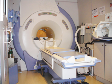
An MRI scanner uses large magnets and a computer to make images. It is used to view the prostate and other organs and tissues nearby. An MRI makes very clear images.
For this test, you lie still on a table as it passes through a tube-like scanner. The scanner sends a type of radio wave at the area of your body to be viewed. A computer puts together the data to create a detailed image of the inside of your body. The technician may use insert a rectal probe to get even better images. If a probe is used, you may have some discomfort.
When the scanner is working, it can be very loud. You may be given earplugs or headphones to wear. The scanner is a small space. If you are uncomfortable in small spaces, you may be given a sedative. Take this before the test. Some hospitals and clinics have open MRI scanners. These are less confining. A 2-way intercom will let you talk to the technician during the test.
Lymph node biopsy
A biopsy is the removal of small pieces of tissue to test. The small pieces of tissue are looked at with a microscope. A lymph node biopsy is done to see if cancer cells have spread to the lymph nodes. These are small collections of immune cells located all around the body. Prostate cancer may spread to nearby lymph nodes.
Lymph node biopsies are not that common in prostate cancer. A biopsy may be done if tests show that you may have cancer in your lymph nodes or other areas of your body. Or a biopsy may be done if scans show that lymph nodes are larger than normal. Treatment for prostate cancer is different if it has spread to the lymph nodes or other areas of your body.
If a biopsy is needed, one or more lymph nodes are removed during surgery (or a less-invasive procedure):
The biopsy may be done at the same time as other surgery for prostate cancer.
The biopsy may also be done as laparoscopic surgery. This uses a few very small cuts (incisions) in the lower belly (abdomen). This procedure is not often used for prostate cancer.
A fine-needle aspiration (FNA) may also be done. A local anesthetic is given. With the help of a CT scan, the lymph nodes are found. A needle is put into the lower abdomen and into the lymph node. A small sample is taken.
A pathologist looks at the tissue with a microscope. A pathologist is a doctor who has special training in examining cells and tissues.
Working with your healthcare provider
Your healthcare provider will talk with you about which tests you'll have. Make sure to get ready for the tests as instructed. Ask questions and talk about any concerns you have.
Updated:
October 15, 2017
Reviewed By:
Alteri, Rick, MD,Gersten, Todd, MD