Legg-Calve-Perthes disease
Legg-Calve-Perthes disease
Natural Standard Monograph, Copyright © 2013 (www.naturalstandard.com). Commercial distribution prohibited. This monograph is intended for informational purposes only, and should not be interpreted as specific medical advice. You should consult with a qualified healthcare provider before making decisions about therapies and/or health conditions.
Related Terms
Anti-inflammatory, arthrogram, arthrography, avascular, avascular hip necrosis, bilateral, bone scans, Chandler disease, femoral head, genitourinary tract, hip necrosis, inguinal canal, inguinal hernia, ischemic, magnetic resonance imaging, MRI, musculoskeletal, necrosis, osteoarthritis, X-rays.
Background
Legg-Calve-Perthes disease is a temporary condition in children in which the ball-shaped head of the thigh bone (femur), also known as the femoral head, loses its blood supply. Hence, the femoral head collapses. The body will absorb the dead bone cells and replace them with new bone cells, eventually reshaping the femoral head of the thigh bone. The femur is the large bone in the thigh, and the femoral head is the rounded ball at the end of the bone that fits into the hip socket. Legg-Calve-Perthes disease causes the hip joint to become painful and stiff for a short period of time.
Legg-Calve-Perthes disease is also known as avascular necrosis or Chandler disease in adults. Avascular necrosis is a disease resulting from the temporary or permanent loss of the blood supply to the bones. Without blood, the bone tissue dies and causes the bone to collapse. Avascular necrosis usually affects people between 30-50 years of age. This monograph pertains to Legg-Calve-Perthes disease in children.
Approximately one in 1,200 children younger than 15 years is affected, usually aged two to 12 years. Legg-Calve-Perthes disease typically affects one hip, but sometimes it develops in both hips. The disease affects both joints in 10-20% of children. When both hips are involved, they are usually affected one after the other and not at the same time. A family history is present in 6% of patients.
Legg-Calve-Perthes disease goes through four phases of changes that affect the head of the femur.
Phase 1: Blood supply is absent to the femoral head and the hip joint becomes inflamed, stiff, and painful. Portions of the bone turn into dead tissue. The ball of the thigh bone becomes less round in appearance on x-rays. This phase can last from several months up to one year. The individual can still walk.
Phase 2: The body cleans up the dead bone cells using the immune system and replaces them with new, healthier bone cells. The femoral head begins to remodel into a round shape again. The joint is still irritated and painful. This phase can last from one to three years.
Phase 3: The femoral head continues to model itself back into a round shape with new bone. This phase lasts for one to three years.
Phase 4: Permanent bone cells replace the new bone cells. This last phase can last a few years to complete the healing process.
The long-term outlook for Legg-Calve-Perthes disease is often good, especially for children who develop the condition very young. The younger the child, the more time there is to reshape the affected hip bone.
Causes and Risk Factors
The underlying cause of Legg-Calve-Perthes disease is not clear. It some cases, it may be due to injury or abnormal blood clotting, which can lead to a lack of blood flow to the bone.
Certain risk factors have been identified in children, including sex, socioeconomic group, and the presence of an inguinal hernia and genitourinary tract complications. An inguinal hernia is an intestinal bulge through a weak spot in the inguinal canal, which is a triangle-shaped opening between layers of abdominal muscle near the groin. More specifically, boys are affected three to five times more often than girls. The incidence increases in low socioeconomic groups and in children with a low birth weight.
The diagnosis of Legg-Calve-Perthes disease must be made as early as possible. Caretakers can watch for signs of decreased mobility, pain, or other complaints in the hip area. Individuals who appear to have the condition after the age of eight years have a poor outcome. Girls have an outcome worse than that of boys, and girls usually have a more severe form of the disease, in part due to hormonal changes as they age.
Ethnicity: Legg-Calve-Perthes disease is most common in Caucasians; it rarely occurs in African Americans.
Gender: The male-to-female ratio is 3-5:1.
Age: Individuals with Legg-Calve-Perthes disease are typically children aged two to 12 years. The mean individual age at presentation is seven years.
The disease may be more likely in physically active children who are small for their age and those who are exposed to secondhand smoke. Smoking causes negative effects on the cardiovascular system and may lead to a decrease in blood supply to the bone.
Signs and Symptoms
The child typically complains of pain in the hip that is made worse by activity. Sometimes, they will also experience pain in their thigh or knee area. The child usually walks with a limp and finds that rest will alleviate the pain.
Limping is often the earliest sign of Legg-Calve-Perthes disease. Pain or stiffness in the hip, groin or knee is possible as well. For some children, the affected leg becomes shorter due to bone collapse.
The symptoms of Legg-Calve-Perthes disease may resemble other conditions or medical problems of the hip, such as osteoarthritis, and may include pain, stiffness, and decreased mobility.
Complications
Legg-Calve-Perthes disease may cause a permanently deformed hip joint, especially if the condition develops after ages six to eight. Severe cases of Legg-Calve-Perthes disease may increase the risk of osteoarthritis and osteoporosis as an adult.
Diagnosis
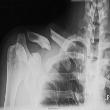
Diagnosis of Legg-Calve-Perthes disease is often based on a child's signs and symptoms, a physical exam, and imaging studies. The imaging studies that a doctor may recommend include X-rays, magnetic resonance imaging (MRI), or bone scans to detect changes in the child's bones. Sometimes Legg-Calve-Perthes disease is detected by accident during an X-ray done for other reasons.
In addition to a complete medical history and physical examination, diagnostic procedures for Legg-Calve-Perthes disease may include:
X-rays: X-ray is a diagnostic test that uses invisible electromagnetic energy beams to produce images of internal tissues, bones, and organs onto film. X-rays are often the first test performed if an individual has symptoms of Legg-Calve-Perthes disease. If the individual has this condition, the X-ray images will often change in the femoral head.
Bone scan: A bone scan locates problems (such as a fracture or osteoporosis) in the spine. A chemical called a radioactive tracer is injected into the child and after several hours, a gamma camera picture will reveal bone undergoing rapid changes where large amounts of tracer accumulate.
Magnetic resonance imaging (MRI): Magnetic resonance imaging (MRI) tests use powerful magnets to produce images on a computer screen and film. Magnetic resonance imaging (MRI) scan provides clear images of musculoskeletal conditions. An MRI is conducted in a small, confined area, and some individuals may find this uncomfortable. Some individuals may have to be sedated using a mild sedative such as alprazolam (Xanax®) or lorazepam (Ativan®). If the individual is sedated, transportation should be organized using a family member or friend.
Arthrograms: An arthrogram is an X-ray exam of a joint after the injection of a dye-like contrast material (non-radioactive) and/or air to outline the soft tissue and joint structures on the pictures. Arthrography is considered extremely safe since reactions to the contrast media are rare.
Blood tests: Blood tests may also be performed in order to determine blood levels of vitamins and minerals, such as calcium, vitamin D, and boron.
Treatment
If the child is diagnosed with Legg-Calve-Perthes disease, he or she may be referred to a special type of doctor called a pediatric orthopedic specialist, for treatment.
The more severe the case, the greater the likelihood that the child may experience limited hip motion, differences in leg lengths, and further hip problems in adulthood.
Specific treatment for Legg-Calve-Perthes disease will be determined by the child's doctor based on: the child's age, overall health, and medical history; the extent of the condition; the child's tolerance for specific medications, procedures, or therapies; expectations for the course of the condition; and the individual's opinion or preference.
The goals of treatment are: to decrease symptoms, to preserve the roundness of the femoral head, and to prevent deformity while the condition runs its course. Treatment options are dependent upon the amount of hip pain, stiffness, and X-ray changes over time, as well as how much of the femoral head has collapsed.
The two most critical factors that determine the outcome are the child's age and how much of the femoral head is affected by this condition. Deterioration of the femoral head can occur at different rates and through different methods.
Anti-inflammatory medications: Over-the-counter medications (OTC), such as ibuprofen (Advil®), can help relieve pain and reduce joint inflammation. These medications are often recommended for months at a time. The dosage may be decreased as the hip begins to heal. Side effect may include mild gastrointestinal upset. These medications should be taken with food.
Physical therapy: Range of motion exercises are exercises that can be done with any part of the body, consisting of a joint pivoting it in a "full range of motion." Rage of motion exercises can help maintain joint mobility. These exercises can be done at home or with the help of a physical therapist.
Crutches: Crutches can ease pain by keeping the child's weight off his or her hip.
Casts, braces, or traction: Temporarily immobilizing the bone can help promote healing. This may be done with leg or hip casts, leg braces, or traction (applying a pulling force to the bone).
Surgery: If a groin muscle has shortened due to excessive limping, it may be surgically released from the bone. After surgery, the affected leg is put in a cast for six to eight weeks to allow the muscle to grow to a more normal length. Sometimes the hip ball must be replaced within the socket. In other cases, the hip socket is repositioned.
Legg-Calve-Perthes disease cannot be prevented. But with appropriate treatment, most children can return to their normal activities within 18 months to two years.
Children with Legg-Calve-Perthes disease may need extra care with personal issues, such as dressing themselves and going to the restroom.
Integrative Therapies
Strong scientific evidence:
Calcium: Calcium is the nutrient consistently found to be the most important for attaining peak bone mass and preventing osteoporosis. Adequate vitamin D intake is required for optimal calcium absorption. Adequate calcium and vitamin D are deemed essential for the prevention of osteoporosis in general, including postmenopausal osteoporosis. Multiple studies of calcium supplementation in the elderly and postmenopausal women have found that high calcium intakes can help reduce the loss of bone density. Studies indicated that bone loss prevention could be achieved in many areas, including ankles, hips, and spine. Although calcium and vitamin D alone are not recommended as the sole treatment of osteoporosis, they are necessary additions to pharmaceutical treatments. Treatment of postmenopausal osteoporosis should only be done under supervision of a qualified healthcare professional.
Avoid if allergic or hypersensitive to calcium or lactose. High doses taken by mouth may cause kidney stones. Avoid with high levels of calcium in the blood, high levels of calcium in urine, hyperparathyroidism (overgrowth of the parathyroid glands), bone tumors, digitalis toxicity, ventricular fibrillation (rapid, irregular twitching of heart muscle), kidney stones, kidney disease, or sarcoidosis (inflammatory disease). Calcium supplements made from dolomite, oyster shells, or bone meal may contain unacceptable levels of lead. Use cautiously with achlorhydria or irregular heartbeat. Talk to a healthcare provider to determine appropriate dosing during pregnancy and breastfeeding.
Vitamin D: Adults with severe vitamin D deficiency lose bone mineral content ("hypomineralization") and experience bone pain, muscle weakness, and osteomalacia (soft bones). Treatment for osteomalacia depends on the underlying cause of the disease and often includes pain control and orthopedic surgical intervention, as well as vitamin D and phosphate binding agents.
Avoid if allergic or hypersensitive to vitamin D or any of its components. Vitamin D is generally well-tolerated in recommended doses; doses higher than recommended may cause toxic effects. Use cautiously with hyperparathyroidism (overactive thyroid), kidney disease, sarcoidosis, tuberculosis, and histoplasmosis. Vitamin D is safe in pregnant and breastfeeding women when taken in recommended doses.
Good scientific evidence:
Vitamin D: Without sufficient vitamin D, inadequate calcium is absorbed and the resulting elevated parathyroid (PTH) secretion causes increased bone resorption. This may weaken bones and increase the risk of fracture. Vitamin D supplementation has been shown to slow osteoporosis and reduce fracture, particularly when taken with calcium.
Avoid if allergic or hypersensitive to vitamin D or any of its components. Vitamin D is generally well-tolerated in recommended doses; doses higher than recommended may cause toxic effects. Use cautiously with hyperparathyroidism (overactive thyroid), kidney disease, sarcoidosis, tuberculosis, and histoplasmosis. Vitamin D is safe in pregnant and breastfeeding women when taken in recommended doses.
Unclear or conflicting scientific evidence:
Black tea: Preliminary research suggests that chronic use of black tea may improve bone mineral density (BMD) in older women. Better research is needed to more clearly determine the effects of black tea for osteoporosis prevention.
Avoid if allergic or hypersensitive to caffeine or tannins. Skin rash and hives have been reported with caffeine ingestion. Use caution with diabetes. Use caution if pregnant. Heavy caffeine intake during pregnancy may increase the risk of SIDS (sudden infant death syndrome). Very high doses of caffeine have been linked with birth defects. Caffeine is transferred into breast milk. Caffeine ingestion by infants can lead to sleep disturbances and insomnia. Infants nursing from mothers consuming greater than 500 milligrams of caffeine daily have been reported to experience tremors and heart rhythm abnormalities. Tea consumption by infants has been linked to anemia, decreased iron metabolism and irritability.
Boron: Animal and preliminary human studies report that boron may play a role in mineral metabolism, with effects on calcium, phosphorus, and vitamin D. However, research of bone mineral density in women taking boron supplements does not clearly demonstrate benefits in osteoporosis. Additional study is needed before a firm conclusion can be drawn.
Avoid if allergic or sensitive to boron, boric acid, borax, citrate, aspartate or glycinate. Avoid with history of diabetes, seizure disorder, kidney disease, liver disease, depression, anxiety, high blood pressure, skin rash, anemia, asthma, or chronic obstructive pulmonary disease (COPD). Avoid with hormone-sensitive conditions like breast cancer or prostate cancer. Avoid if pregnant or breastfeeding.
Calcium: Calcium supplementation above the normal daily dietary intake has not been shown to reduce stress fractures. Further studies are needed to better determine the role of calcium in bone stress injury prevention.
Rickets and osteomalacia (bone softening) are commonly thought of as diseases due to vitamin D deficiency; however, calcium deficiency may also be another cause in sunny areas of the world where vitamin D deficiency would not be expected. Calcium gluconate is used as an adjuvant in the treatment of rickets and osteomalacia, as well as a single therapeutic agent in non-vitamin D deficient rickets. Research continues into to the importance of calcium alone in the treatment and prevention of this condition. Treatment of rickets and osteomalacia should only be done under the supervision of a qualified healthcare professional.
Avoid if allergic or hypersensitive to calcium or lactose. High doses taken by mouth may cause kidney stones. Avoid with high levels of calcium in the blood, high levels of calcium in urine, hyperparathyroidism (overgrowth of the parathyroid glands), bone tumors, digitalis toxicity, ventricular fibrillation (rapid, irregular twitching of heart muscle), kidney stones, kidney disease, or sarcoidosis (inflammatory disease). Calcium supplements made from dolomite, oyster shells, or bone meal may contain unacceptable levels of lead. Use cautiously with achlorhydria or irregular heartbeat. Talk to a healthcare provider to determine appropriate dosing during pregnancy and breastfeeding.
Copper: Supplementation with copper may be helpful in the treatment and/or prevention of osteoporosis, although early human evidence is conflicting. Further research is needed before clear conclusions can be drawn.
Avoid if allergic to copper. Avoid copper supplements during the early phase of recovery from diarrhea. Avoid with hypercupremia. Avoid with genetic disorders affecting copper metabolism (e.g. Wilson's disease, Indian childhood cirrhosis, or idiopathic copper toxicosis). Avoid with HIV/AIDS. Use cautiously with water containing copper concentrations greater than 6 milligrams/liter. Use cautiously with anemia, arthralgias, or myalgias. Use cautiously if taking birth control pills. Use cautiously if at risk for selenium deficiency. Doses that do not exceed the recommended dietary allowance appear to be safe during pregnancy and breastfeeding.
Creatine: Creatine is an amino acid that is found in the muscles. Early studies examining the effect of creatine in aging suggest that creatine may increase bone density when combined with resistance training. Further studies in which creatine alone is compared with placebo are needed.
Avoid if allergic or hypersensitive to creatine. Early research suggests that creatine may reduce muscle cramps that are often associated with hemodialysis. However, further studies are needed to confirm this claim. Avoid if taking diuretics (e.g. hydrochlorothiazide or furosemide). Use cautiously with asthma, diabetes, gout, kidney disease, liver disease, muscle problems, stroke, or with a history of these conditions. Avoid dehydration. Avoid if pregnant or breastfeeding.
DHEA: DHEA (dehydroepiandrosterone) is a hormone made in the human body and secreted by the adrenal gland. The ability of DHEA to increase bone density is under investigation. Effects are not clear at this time.
Avoid if allergic to DHEA. Avoid with a history of seizures. Use with caution in adrenal or thyroid disorders or with anticoagulants, or drugs, herbs or supplements for diabetes, heart disease, seizure, or stroke. Stop use two weeks before surgery/dental/diagnostic procedures with bleeding risk, and do not use immediately after these procedures. Avoid if pregnant or breastfeeding.
Evening primrose oil: Primrose oil has been suggested as a possible treatment for osteoporosis. Well-designed human trials are needed before primrose oil can be recommended for osteoporosis therapy.
Avoid if allergic to plants in the Onagraceae family (willow's herb, enchanter's nightshade) or gamma-linolenic acid. Avoid with seizure disorders. Use cautiously with mental illness drugs. Stop use two weeks before surgery with anesthesia. Avoid if pregnant or breastfeeding.
Gamma linolenic acid (GLA): Gamma linolenic acid (GLA) is a dietary omega-6 fatty acid found in many plant oil extracts. Some clinical evidence suggests that GLA and eicosapentaenoic acid (EPA) enhance the effects of calcium supplementation for osteoporosis. More clinical studies are required to better determine effectiveness.
Use cautiously with drugs that increase the risk of bleeding like anticoagulants and anti-platelet drugs. Avoid if pregnant or breastfeeding.
Horsetail: Silicon may be beneficial for bone strengthening. Because horsetail (Equisetum arvense) contains silicon, it has been suggested as a possible natural treatment for osteoporosis. Preliminary human study reports benefits, but more detailed research is needed before a firm recommendation can be made. People with osteoporosis should speak with a qualified healthcare provider about possible treatment with more proven therapies.
Avoid if allergic or hypersensitive to horsetail or nicotine. Avoid with a history of chronic alcohol abuse, malnutrition, and kidney disorders. Use cautiously with abnormal heart rhythms, diabetes, gout, neurological disorders, and osteoporosis. Avoid in children. Avoid if pregnant or breastfeeding.
Phosphates, phosphorus: Early research shows that high amounts of phosphorus may have negative effects on bone density. This is because phosphorus decreases bone formation and increases bone resorption. Additional study is needed in this area.
Avoid if allergic or hypersensitive to any ingredients in phosphorus/phosphate preparations. Use phosphorus/phosphate salts cautiously with kidney or liver disease, heart failure, unstable angina (chest pain), recent heart surgery, hyperphosphatemia (high phosphate blood level), hypocalcemia (low calcium blood level), hypokalemia (low potassium blood level), hypernatremia (high sodium blood level), Addison's disease, intestinal obstruction or ileus, bowel perforation, severe chronic constipation, acute colitis, toxic megacolon, hypomotility syndrome, hypothyroidism, scleroderma, or gastric retention. Avoid sodium phosphate enemas with congenital or abnormalities of the intestine. Too much phosphorus may cause serious or life-threatening toxicity.
Physical therapy: Supervised or home-based physical therapy has been used in combination with resistance and endurance training in physically frail elderly women taking hormone replacement therapy to improve bone density. Although early study is promising, more studies are needed in this area.
Not all physical therapy programs are suited for everyone, and patients should discuss their medical history with a qualified healthcare professional before beginning any treatments. Physical therapy may aggravate pre-existing conditions. Persistent pain and fractures of unknown origin have been reported. Physical therapy may increase the duration of pain or cause limitation of motion. Pain and anxiety may occur during the rehabilitation of patients with burns. Both morning stiffness and bone erosion have been reported in the literature although causality is unclear. Erectile dysfunction has also been reported. Physical therapy has been used in pregnancy and although reports of major adverse effects are lacking in the available literature, caution is advised nonetheless. All therapies during pregnancy and breastfeeding should be discussed with a licensed obstetrician/gynecologist before initiation.
Red clover: Red clover (Trifolium pratense) is a legume, which like soy, contains "phytoestrogens" (plant-based chemicals that are similar to estrogen, and may act in the body like estrogen or may actually block the effects of estrogen). It is not clear if red clover isoflavones have beneficial effects on bone density. Most studies of isoflavones in this area have looked at soy, which contains different amounts of isoflavones, as well as other non-isoflavone ingredients. More research is needed to better understand the effects of red clover on osteoporosis.
Avoid if allergic to red clover or other isoflavones. Use caution with hormone replacement therapy (HRT) or birth control pills. Use caution with history of a bleeding disorder, breast cancer, or endometrial cancer. Use caution with drugs that thin the blood. Avoid if pregnant or breastfeeding.
Soy: It has been theorized that phytoestrogens in soy (such as isoflavones) may reduce the risk of osteoporosis. However, more research is needed before a conclusion can be made.
Avoid if allergic to soy. Breathing problems and rash may occur in sensitive people. Soy, as a part of the regular diet, is traditionally considered to be safe during pregnancy and breastfeeding, but there is limited scientific data. The effects of high doses of soy or soy isoflavones in humans are not clear, and therefore are not recommended. People who experience intestinal irritation (colitis) from cow's milk may experience intestinal damage or diarrhea from soy. It is not known if soy or soy isoflavones share the same side effects as estrogens, like increased risk of blood clots. The use of soy is often discouraged in patients with hormone-sensitive cancers, such as breast, ovarian, or uterine cancer. Other hormone-sensitive conditions such as endometriosis may also be worsened. Patients taking blood-thinning drugs like warfarin should check with a doctor and pharmacist before taking soy supplementation.
Tai chi: Tai chi is a system of movements and positions believed to have developed in 12th Century China. Tai chi techniques aim to address the body and mind as an interconnected system and are traditionally believed to have mental and physical health benefits to improve posture, balance, flexibility, and strength. Preliminary research suggests that tai chi may be beneficial in delaying early bone loss in postmenopausal women and preventing osteoporosis. Additional evidence and long-term follow-up is needed to confirm these results.
Avoid with severe osteoporosis or joint problems, acute back pain, sprains, or fractures. Avoid during active infections, right after a meal, or when very tired. Some believe that visualization of energy flow below the waist during menstruation may increase menstrual bleeding. Straining downwards or holding low postures should be avoided during pregnancy, and by people with inguinal hernias. Some tai chi practitioners believe that practicing for too long or using too much intention may direct the flow of chi (qi) inappropriately, possibly resulting in physical or emotional illness. Tai chi should not be used as a substitute for more proven therapies for potentially serious conditions. Advancing too quickly while studying tai chi may increase the risk of injury.
Vitamin K: Vitamin K appears to prevent bone resorption, and adequate dietary intake is likely necessary to prevent excess bone loss and for osteoporosis prevention. Elderly or institutionalized patients may be at particular risk and adequate intake of vitamin K-rich foods should be maintained. Unless patients have demonstrated vitamin K deficiency, there is no evidence that additional vitamin K supplementation is helpful.
Avoid if allergic or hypersensitive to vitamin K. Injection into the muscle or vein should only be done by a healthcare professional; many serious side effects have occurred after injection. Menadiol (type of vitamin K that is not available in the United States) should be avoided with glucose-6-phosphate dehydrogenase deficiency. Conditions that interfere with absorption of ingested vitamin K may lead to deficiency, including short gut, cystic fibrosis, malabsorption (various causes), pancreas or gall bladder disease, persistent diarrhea, sprue, or ulcerative colitis. Avoid if pregnant. Use cautiously if breastfeeding.
Prevention
Sunlight: Healthcare professionals recommend sun exposure of 15 minutes a day to the hands and face to help the body make vitamin D. Vitamin D helps calcium be absorbed and used by the body. Avoid overexposure to the sun. Use sunscreen on children when exposed to intense sunlight.
Healthy diet: Being underweight is a risk factor for bone loss. Eating a healthy diet and staying within a healthy weight is important.
A high protein diet increases calcium excretion and may increase the calcium needs for the body. Fiber, oxalates (in rhubarb, spinach, beets, celery, greens, berries, nuts, tea, cocoa), and high zinc foods (such as oysters and red meats) decrease absorption, requiring more calcium as a dietary supplement. Healthcare professionals recommend following the Dietary Guidelines for Americans published by the U.S. Department of Agriculture in 2005.
Eliminating fall hazards: If a child has bone loss, it is important not only to help prevent further bone loss, but also to prevent a fracture. Eliminating hazards in the house that can increase the risk of falling is important. Wearing sturdy shoes is important. Caretakers should not hesitate to take a child with Legg-Calve-Perthes disease to the doctor is the child is having hip or leg pain after a fall.
Author Information
This information has been edited and peer-reviewed by contributors to the Natural Standard Research Collaboration (www.naturalstandard.com).
Bibliography
Natural Standard developed the above evidence-based information based on a thorough systematic review of the available scientific articles. For comprehensive information about alternative and complementary therapies on the professional level, go to www.naturalstandard.com. Selected references are listed below.
American Academy of Family Physicians. http://familydoctor.org.
Brech GC, Guarnieiro R. Evaluation of physiotherapy in the treatment of Legg-Calve-Perthes disease. Clinics. 2006;61(6):521-8. View Abstract
Eijer H, Berg RP, Haverkamp D, et al. Hip deformity in symptomatic adult Perthes' disease. Acta Orthop Belg. 2006;72(6):683-92. View Abstract
Nathan Sambandam S, Gul A, Shankar R, et al. Reliability of radiological classifications used in Legg-Calve-Perthes disease. J Pediatr Orthop B. 2006;15(4):267-70. View Abstract.
National Institute of Arthritis and Musculoskeletal and Skin Diseases (NIAMS). www.niams.nih.gov.
National Institute on Aging. www.nia.nih.gov.
Natural Standard: The Authority on Integrative Medicine. www.naturalstandard.com.
Poul J. Diagnosis of Legg-Calve-Perthes disease. Ortop Traumatol Rehabil. 2004;6(5):604-6. View Abstract
Zarzycka M, Zarzycki D, Kacki W, et al. Long-term results of conservative treatment in Perthes' disease. Ortop Traumatol Rehabil. 2004;6(5):595-603. View Abstract
Copyright © 2013 Natural Standard (www.naturalstandard.com)
The information in this monograph is intended for informational purposes only, and is meant to help users better understand health concerns. Information is based on review of scientific research data, historical practice patterns, and clinical experience. This information should not be interpreted as specific medical advice. Users should consult with a qualified healthcare provider for specific questions regarding therapies, diagnosis and/or health conditions, prior to making therapeutic decisions.
Updated:
March 22, 2017