Facioscapulohumeral muscular dystrophy
Facioscapulohumeral muscular dystrophy
Natural Standard Monograph, Copyright © 2013 (www.naturalstandard.com). Commercial distribution prohibited. This monograph is intended for informational purposes only, and should not be interpreted as specific medical advice. You should consult with a qualified healthcare provider before making decisions about therapies and/or health conditions.
Related Terms
Bilateral sensorineural hearing loss, chromosome 4-linked FSHD, Coats' disease, facio-scapulo-humeral muscular dystrophy, fascioscapulohumeral muscular dystrophy, FSH, FSHD, FSHD type 1A, FSHD type 1B, FSHMD, hypercarbic respiratory insufficiency, IFSHD, infantile FSHD, Landouzy-Dejerine muscular dystrophy, position effect retinal telangiectasis, scapulohumeral muscular dystrophy, muscle wasting, neuromuscular, non-chromosome 4-linked FSHD, somatic mosaicism, subtelomeric repeat exchange.
Background
Facioscapulohumeral muscular dystrophy (FSHD) is a disease that gradually causes muscles to deteriorate. Muscular dystrophies in general are hereditary muscle diseases that cause progressive muscle weakness. FSHD predominantly affects the upper body, unlike Duchenne muscular dystrophy and Becker muscular dystrophy, which affect the lower body. Doctors Landouzy and Dejerine first described FSHD in 1885 and, as a result, the disease is sometimes called Landouzy-Dejerine muscular dystrophy.
The major symptom of FSHD is the gradual weakening and loss of skeletal muscles. The usual location of these weaknesses give the disease its name: face (facio), shoulder girdle (scapulo), and upper arms (humeral). There is wide variability in the age at which symptoms first appear and the severity of the disease, which varies even within affected members of the same family.
FSHD is caused by a genetic defect or mutation in a person's DNA (deoxyribonucleic acid). DNA is packaged on larger structures called chromosomes. Humans have 23 pairs of chromosomes, including 22 pairs of autosomes and one pair of sex chromosomes. Almost all cases of FSHD are associated with a deletion, or a piece of missing DNA, from chromosome 4. This region of deleted DNA does not belong to any known genes, but is believed to be involved in the regulation of other genes.
FSHD has been classified into three types: FSHD type 1A (chromosome 4-linked FSHD), FSHD type 1B (non-chromosome 4-linked FSHD), and infantile FSHD (IFSHD). The symptoms are the same for all types. The difference between the types depends on the type of mutation that causes the disease. More than 98% of cases are of the FSHD type 1A.
Because FSHD is inherited, the only known risk factor is a family history of the disorder. It affects men and women equally. FSHD is distributed worldwide and does not occur to a greater degree in any racial or ethnic group or geographic location.
Although there is currently no known cure for FSHD, many treatments are available to improve symptoms and maximize the quality of life. With proper treatment, many individuals with FSHD have normal life expectancies.
Risk Factors
General: Because facioscapulohumeral muscular dystrophy (FSHD) is inherited, the only known risk factor is a family history of the disorder. It affects men and women equally. FSHD does not occur at a higher rate in any racial or ethnic group or geographic location.
Autosomal dominant inheritance: FSHD is inherited as an autosomal dominant trait, meaning that a child has to inherit only one copy of the defective (mutated) gene to have the disease. Individuals inherit two copies of most genes, one from the mother and one from the father. If one parent has FSHD, there is a 50% chance that each of his or her children will have the disorder. If both parents have the disorder, there is a 75% chance that each child will have FSHD. The type (FSHD 1A, FSHD 1B, or infantile FSHD) of FSHD inherited by the child is always the same as that of the affected parent.
Random occurrence: A small percentage of cases of FSHD occur in individuals with a family history of the disease. This may occur because of a random, or sporadic, mutation in the egg, sperm, or developing embryo. When FSHD occurs in this manner, it can then be transmitted to future generations in an autosomal dominant manner.
Incidence: The most common form of FSHD is FSHD type 1A, which is estimated to occur in approximately one in 20,000 births throughout the world. The other two types, FSHD type 1B and infantile FSHD, are very rare and are believed to account for less than 2% of all FSHD cases.
Causes
General: Almost all cases of facioscapulohumeral muscular dystrophy (FSHD) are associated with a deletion, or a piece of missing DNA, from chromosome 4. This region of deleted DNA does not belong to any known genes but is believed to be involved in the regulation of other genes. The deleted region of DNA is composed of a number of copies of a piece of DNA called the "D4Z4." A normal chromosome has between 11 and 150 copies of D4Z4. Some people inherit more copies of D4Z4 than others. Individuals with 11 or fewer copies of D4Z4 are considered to be at risk for developing FSHD.
Autosomal dominant inheritance: FSHD is inherited as an autosomal dominant trait, meaning that a child has to inherit only one copy of the mutated gene to have the disease. Individuals inherit two copies of most genes, one from the mother and one from the father. If one parent has FSHD, there is a 50% chance that each of his or her children will have the disorder. If both parents have the disorder, there is a 75% chance that each child will have FSHD. The type (FSHD 1A, FSHD 1B, or infantile FSHD) of FSHD inherited by the child is always the same as that of the affected parent.
Random occurrence: A small percentage of people with FSHD do not inherit the genetic mutation or defect associated with the disease. This may occur because of a random, or sporadic, mutation in the egg, sperm, or developing embryo. When FSHD occurs in this manner, it can then be transmitted to future generations in an autosomal dominant manner.
Signs and Symptoms
General: The major symptom of facioscapulohumeral muscular dystrophy (FSHD) is the gradual weakening and loss of skeletal muscles. The usual location of these weaknesses gives the disease its name: face (facio), shoulder girdle (scapulo), and upper arms (humeral). There is wide variability in the age at which symptoms first appear and disease severity, which varies even within affected members of the same family.
Symptoms of FSHD usually occur between 10 and 20 years of age. In some cases, however, symptoms never develop even when the individual has the mutation associated with FSHD. In most cases, muscle weakening starts in the face and slowly progresses to the shoulder and upper arm muscles and then down to the stomach and foot muscles. Foot drop, which is an inability to flex the foot upward, and foot weakness are common symptoms. For unknown reasons, most FSHD patients develop unbalanced weaknesses, where one side of the body is weaker than the other.
Abdomen: Weakness in the abdominal muscles can cause a bulging abdomen and lumbar lordosis, or inward curving of the lower spine. The lower abdominal muscles are usually weaker than the upper abdominal muscles. This pattern of abdominal muscle weakness is unique to FSHD.
Face: Facial muscle weakness is common and may include eyelid drooping, decreased facial expression, depressed or angry facial expression, and difficulty pronouncing the letters M, B, and P. Early weakness of the muscles of the eye (including the ability to open and close the eye) and mouth (inability to smile, pucker, or whistle) are also common.
Hearing: Patients with FSHD may experience high-tone deafness. This symptom is rare and may lead to hearing loss.
Legs: FSHD can affect muscles around the pelvis, hips, and lower legs, and may interfere with walking as the disorder gets worse.
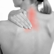
Shoulder: Shoulder muscle weakness causes deformities such as pronounced shoulder blades (scapular winging) and sloping shoulders. FSHD patients may have difficulty raising their arms because of shoulder and arm muscle weakness. Unequal weakening of the upper and lower arm and upper back muscles may occur.
Types of the Disease
General: Facioscapulohumeral muscular dystrophy (FSHD) has been classified into three types: FSHD1A, FSHD1B, and IFSHD. The symptoms are the same for each type.
FSHD1A: The most common form of FSHD is FSHD type 1A, or chromosome 4-linked FSHD. FSHD1A can be detected by genetic testing for missing or deleted DNA from a region of chromosome 4 called D4Z4. Patients missing the most DNA from this region typically have the most severe symptoms, such as an inability to walk.
FSHD1B: FSHD type 1B, or non-chromosome 4-linked FSHD, is much less common than FSHD1A. Researchers are not sure whether it is caused by mutations in the genes that cause FSHD1A or in other genes.
IFSHD: Infantile FSHD (IFSHD) is rare. It is a more severe form of FSHD1A or FSHD1B, in which symptoms occur in early childhood. What makes the disease more severe has not yet been determined. Hearing loss, vision problems, and seizures are associated with IFSHD.
Diagnosis
Physical exam: A physical examination of a patient with facioscapulohumeral muscular dystrophy (FSHD) would reveal weakness of the facial and shoulder muscles. High blood pressure may be noted but is usually mild. An eye exam may show changes in the blood vessels in the back of the eye (retinal telangiectasias).
Creatine kinase (CK) test: Tests that measure an enzyme called creatine kinase (CK, also known as creatine phosphokinase or CPK) in the blood can be used to identify muscle defects. CK is an enzyme that helps carry out a chemical reaction on creatine, a substance used by muscle for energy. Because CK may leak out of damaged muscle cells, CK levels are increased in the blood of patients with FSHD. CK levels may also be increased for other reasons, such as a heart attack or normal exercise. Because normal exercise and diseases that affect the muscles also result in increased levels of serum CK, the serum CK test may not on its own be enough to diagnose a patient with FSHD.
EMG (electromyography): An electromyogram measures electrical impulses in muscle tissue. Muscle weakness can cause these impulses to be diminished in patients with FSHD.
Genetic testing: Genetic testing that measures the size of the D4Z4 deletions on chromosome 4 is the preferred mechanism for diagnosing FSHD. This type of testing is highly accurate but may be expensive.
Muscle biopsy: In an outpatient setting, a small piece of muscle is removed, usually from the arm or leg. This tissue sample is then evaluated with a variety of biochemical tests for FSHD that measure muscle deterioration.
Nerve conduction velocity (NCV): This test measures how fast signals travel from one part of a nerve to another. The nerve signals are measured with surface electrodes similar to those used for an electrocardiogram. Patients with FSHD have slower than normal NCV.
Complications
General: Complications of facioscapulohumeral muscular dystrophy (FSHD) may include decreased mobility, decreased ability to care for oneself, deformities of the face and shoulders, and, rarely, breathing problems. Life expectancy is normal, but up to 20% of affected individuals become severely disabled and eventually must use a wheelchair. Abnormal heart rhythms may occur but tend to be rare.
Hearing loss: Patients with FSHD may experience high-frequency hearing loss caused by nerve damage in both ears. This is more common in infantile FSHD.
Vision: Rarely, FSHD patients have retinal telangiectasias (also called Coats' disease), which is an abnormality of blood vessels in the back of the eye. This symptom occurs in about 1% of cases and is usually associated with infantile FSHD.
Treatment
General: There is currently no known cure for facioscapulohumeral muscular dystrophy (FSHD). Treatment of FSHD aims to reduce symptoms and to improve the individual's quality of life.
Assistive devices: As FSHD progresses and the patient loses mobility, assistive devices such as braces, standing frames, walkers, and wheelchairs may be needed. Braces on the lower legs may also help to lengthen the muscles and prevent contractures, a shortening of the muscle fibers that limit range of motion and prevent mobility. A brace may be prescribed for wear while sleeping to keep the foot from pointing downward and to keep the Achilles tendon stretched. Other devices that may be helpful include a transfer board for moving in and out of the wheelchair, mechanical lifts, shower chairs, and adjustable beds. Scapular bracing may help to stabilize the shoulder.
Medications: Stamulumab, also known as MYO-029, is an experimental myostatin-inhibiting drug for the treatment of muscular dystrophy. Myostatin is a protein that inhibits the growth of muscle tissue. By inhibiting myostatin, Stamulumab stimulates muscle growth.
Albuterol, also known as salbutamol, is known to have muscle-building effects that may be beneficial for FSHD. A human trial evaluating the use of albuterol showed no significant difference in muscle strength at one year in FSHD patients, but lean body mass (fat-free body weight) and grip strength did improve.
Creatine is a molecule that has been used as a muscle performance enhancer by athletes and it may be helpful in muscular dystrophies, including FSHD. Creatine is naturally synthesized in the human body from amino acids primarily in the kidney and liver and is transported in the blood for use by muscles. Further research of creatine supplementation for muscular dystrophy is needed before a recommendation can be made.
Occupational therapy: Occupational therapy may be used to help individuals with FSHD learn to use new devices. Occupational therapy aims to improve function in specific activities, such as aspects of self care, driving, and using a computer.
Physical therapy: Physical therapy may help individuals with FSHD maintain muscle strength. Because inactivity may worsen muscle conditioning in people with FSHD, activity is encouraged. The goals of physical therapy are to allow greater motion in the joints and to maintain muscle strength and tone. Swimming and water exercises may help individuals with FSHD keep muscles strong and toned without causing stress. Exercise should not be done to the point of exhaustion.
Psychological support: Because of the stress experienced by individuals who have degenerative diseases, psychotherapy or a support group may be helpful for patients and families with FSHD. Muscular dystrophy support groups, for example, provide a forum in which members can share experiences, problems, and coping strategies. Individuals with learning disabilities can also be evaluated by a mental health professional. Special exercises and educational therapies may be prescribed.
Respiratory support: If breathing problems become severe, respiratory assistance may be needed. A special mask worn while sleeping can deliver positive airway pressure. As breathing deteriorates, a cough assistance device can help the patient cough and keep the lungs clear.
Speech therapy: If speech and swallowing are affected by muscle wasting of FSHD, a speech therapist may be helpful. Recommendations may include avoiding certain foods and eating or drinking in positions that can reduce the chances of choking or inhaling food or liquid into the lungs.
Surgery: A shoulder blade (scapular) fusion may be performed to permanently fuse the shoulder blade to the rest of the bones in the chest area (thorax).
Integrative Therapies
Note: Currently, there are limited scientific data on the use of integrative therapies for the treatment or prevention of facioscapulohumeral muscular dystrophy (FSHD). While the use of these therapies to treat FSHD has not been well studied, these therapies may potentially be effective in treating symptoms of muscle weakness. The integrative therapies listed below should be used only under the supervision of a qualified healthcare provider and should not be used in replacement of other proven therapies.
Good scientific evidence:
Vitamin D: Vitamin D deficiency has been associated with muscle weakness and pain in both adults and children. Limited research has reported vitamin D deficiency in patients with low back pain, and supplementation may lead to pain reduction in many patients. Avoid if allergic or hypersensitive to vitamin D or any of its components. Vitamin D is generally well tolerated in recommended doses. Doses higher than recommended may cause toxic effects. Individuals with hyperparathyroidism (overactive thyroid), kidney disease, sarcoidosis (immune system disorder), tuberculosis, or histoplasmosis (fungal disease) are at a higher risk of experiencing toxic effects. Vitamin D is generally considered safe for pregnant women. It may be necessary to give infants vitamin D supplements along with breast milk. The recommended intake of vitamin D for normal infants, children, and adolescents is 200 International Units (IU) daily.
Unclear or conflicting scientific evidence:
Coenzyme Q10: Coenzyme Q10 (CoQ10) is produced by the human body and is necessary for the basic functioning of cells. CoQ10 levels are reported to decrease with age and to be low in patients with some chronic diseases such as muscular dystrophies. Early studies in patients with muscular dystrophy taking CoQ10 supplements describe improvements in exercise capacity, heart function, and overall quality of life. Additional research is needed in this area.
Avoid in patients with allergy or hypersensitivity to CoQ10. Although few side effects have been associated with CoQ10, there have been reports of nausea, stomach upset, or rash. Caution is advised in people who have bleeding disorders or who are taking drugs that increase the risk of bleeding such as aspirin and warfarin. Caution is advised in patients with diabetes or hypoglycemia (low blood sugar) and in those taking drugs, herbs, or supplements that affect blood sugar. Use cautiously in patients with liver disease, as large doses of CoQ10 (greater than 300mg per day) may elevate aminotransferase levels. Aminotransferases are important in regulating protein levels. Use cautiously in patients with biliary obstruction or liver disease, as these conditions may increase CoQ10 concentrations. CoQ10 may decrease blood pressure, and caution is advised in patients with low blood pressure or in those taking blood pressure medications. Elevations of liver enzymes have been reported rarely, and caution is advised in people with liver disease or in those taking medications that may harm the liver. CoQ10 may lower blood levels of cholesterol or triglycerides. Based on limited human evidence, thyroid hormone levels may be altered. There is not enough scientific evidence to support the use of CoQ10 during pregnancy or breastfeeding.
Creatine: Creatine is naturally synthesized in the human body from amino acids primarily in the kidney and liver and transported in the blood for use by muscles. Creatine loss is suspected to cause muscle weakness and breakdown in Duchenne muscular dystrophy. Further research of creatine supplementation for muscular dystrophy is needed before a recommendation can be made.
Avoid in patients with allergy or hypersensitivity to creatine. Use of creatine supplements has been associated with symptoms of asthma. There have been rare reports of loss of appetite, stomach upset, diarrhea, or nausea with creatine use. Avoid in patients with liver or kidney disease. Use cautiously in patients with diabetes or low blood sugar. Creatine may cause muscle cramps or muscle breakdown, leading to muscle tears or discomfort. Weight gain and increased body mass may occur. Heat intolerance, fever, dehydration, reduced blood volume, or electrolyte imbalances and resulting seizures may occur. Chronic administration of a large quantity of creatine is reported to increase the production of formaldehyde, which may potentially cause serious unwanted side effects. Creatine may increase the risk of compartment syndrome of the lower leg, a condition characterized by pain in the lower leg associated with inflammation and ischemia (diminished blood flow), which is a potential surgical emergency. Creatine cannot be recommended during pregnancy or breastfeeding due to a lack of safety information.
Fair negative scientific evidence:
Selenium: Early studies suggest that selenium supplementation is not helpful in muscular dystrophy. Avoid if allergic or sensitive to products containing selenium. Avoid with a history of skin cancer. Selenium is generally regarded as safe for pregnant or breastfeeding women. However, animal research reports that large doses of selenium may lead to birth defects.
Prevention
Because facioscapulohumeral muscular dystrophy (FSHD) is inherited, there is currently no known way to prevent the condition.
Genetic counseling: Genetic counseling can help individuals, families, and couples affected by or at risk for FSHD to work through the process of genetic testing for the disease. Genetic counseling also helps individuals, families, and couples planning to have a baby. In vitro fertilization pre-implantation genetic (PGD IVF) tests are available for FSHD1A (chromosome 4-linked FSHD). In PGD IVF, embryos are tested for the defective genes, and only the embryos that are not affected may be implanted. Because FSHD can be detected in a developing fetus, parents may choose whether or not to continue the pregnancy. Genetic counselors help couples consider possible scenarios and alternatives, such as having a child with FSHD, adoption, or artificial insemination.
Prenatal diagnosis: Prenatal testing that measures the size of the D4Z4 deletions on chromosome 4 of the fetus is the preferred mechanism for diagnosing FSHD. This type of testing is highly accurate but may be expensive. The cells for this test can be obtained from chorionic villus sampling, amniocentesis, or from umbilical cord blood. All prenatal tests have a risk of miscarriage.
During amniocentesis, a long, thin needle is inserted through the abdominal wall into the uterus and a small amount of amniotic fluid is removed from the sac surrounding the fetus. The fluid is then analyzed to measure the size of the D4Z4 deletions on chromosome 4. This test is performed after 15 weeks of pregnancy. Some experts estimate that the risk of miscarriage ranges from one in 200 to one in 400 patients. Risk for miscarriage is highest when the procedure is done early in pregnancy, before the two layers of fetal membranes have sealed. A woman's particular risk depends in large part on the skill and experience of the doctor performing the procedure. Some patients may experience minor complications, such as cramping, leaking fluid, or irritation where the needle was inserted.
During chorionic villus sampling (CVS), a small piece of tissue (chorionic villi) is removed from the placenta during early pregnancy. Depending on where the placenta is located, CVS can be performed through the cervix or through the abdomen. The tissue sample is then analyzed to measure the size of the D4Z4 deletions on chromosome 4. This procedure may be performed between the ninth and 14th weeks of pregnancy. The risks of infection or fetal damage are slightly higher with CVS than with amniocentesis. Miscarriage occurs in about two out of 100 women who undergo this procedure. There appears to be an even higher risk of miscarriage with the transcervical CVS technique compared to the transabdominal technique. Other factors that further increase the risks of CVS include having the procedure performed three or more times and having a fetus that is smaller than normal for their gestational age. The physician's skill and experience also play an important role.
Author Information
This information has been edited and peer-reviewed by contributors to the Natural Standard Research Collaboration (www.naturalstandard.com).
Bibliography
Natural Standard developed the above evidence-based information based on a thorough systematic review of the available scientific articles. For comprehensive information about alternative and complementary therapies on the professional level, go to www.naturalstandard.com. Selected references are listed below.
Clapp J, Bolland DJ, Hewitt JE. Genomic analysis of facioscapulohumeral muscular dystrophy. Brief Funct Genomic Proteomic. 2003 Oct;2(3):213-23. View Abstract
Facioscapulohumeral Muscular Dystrophy Society. www.fshsociety.org.
Natural Standard: The Authority on Integrative Medicine. www.naturalstandard.com.
National Center for Biotechnology Information: Online Mendelian Inheritance in Man. www.ncbi.nlm.nih.gov.
Rose MR, Tawil R. Drug treatment for facioscapulohumeral muscular dystrophy. Cochrane Database Syst Rev. 2004;(2):CD002276. View Abstract
Tawil R, Van Der Maarel SM. Facioscapulohumeral muscular dystrophy. Muscle Nerve. 2006 Jul;34(1):1-15. View Abstract
Tupler R, Gabellini D. Molecular basis of facioscapulohumeral muscular dystrophy. Cell Mol Life Sci. 2004 Mar;61(5):557-66. View Abstract
van der Maarel SM, Frants RR, Padberg GW. Facioscapulohumeral muscular dystrophy. Biochim Biophys Acta. 2007 Feb;1772(2):186-94. View Abstract
Copyright © 2013 Natural Standard (www.naturalstandard.com)
The information in this monograph is intended for informational purposes only, and is meant to help users better understand health concerns. Information is based on review of scientific research data, historical practice patterns, and clinical experience. This information should not be interpreted as specific medical advice. Users should consult with a qualified healthcare provider for specific questions regarding therapies, diagnosis and/or health conditions, prior to making therapeutic decisions.
Updated:
March 22, 2017