Diagnosing Interstitial Lung Disease
Diagnosing Interstitial Lung Disease
Your health history
You may be asked some of these questions:
Do you have shortness of breath?
What medicines do you take?
Have you breathed in any harmful substances over a long period of time? This could include molds, chemicals, asbestos, silica, or coal dust.
Do you smoke?
Have you had radiation therapy? This could be to treat breast cancer, for example.
Have you had lung infections?
Is there a family history of lung disease or connective tissue disorders?
Tests
You may need routine blood tests and a chest X-ray. Other tests may include:
ECG (electrocardiogram). This test checks your heart rhythm. It helps rule out heart disease as the cause of your shortness of breath.
CT scan. This test makes a more detailed X-ray image of your lungs.
Pulse oximetry. This test uses a small device placed on a fingertip. It measures the level of oxygen in your blood at rest and with exercise.
Arterial blood gas. This test measures the level of oxygen in your blood. The healthcare provider take a blood sample from an artery, usually at your wrist.
Cardiopulmonary stress test. This test checks how well your heart and lungs work when you are active. You usually ride a stationary bike or walk on a treadmill.
Bronchoscopy. This test allows the doctor to look inside your airways with a thin, lighted tube (bronchoscope). The bronchoscope is passed through your mouth and throat and into your lungs. The doctor may pass tools through the bronchoscope to remove small samples of your lung tissue. This is called a biopsy. The tissue samples can help check for damage or inflammation in your lungs.
Surgical lung biopsy. This test removes samples of your lung tissue through one or more cuts (incisions) made between your ribs or in your chest. The tissue samples are often larger than those taken with bronchoscopy. They may provide more accurate diagnosis of your lung problem.
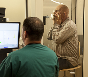
Pulmonary function tests (PFTs)
PFTs are done in the healthcare provider’s office or in the hospital lab. You may be tested both before and after taking medicine. And you may have to repeat the tests from time to time during your treatment. Your healthcare provider may order:
Spirometry. This test measures how much air you can breathe out after a deep breath (forced vital capacity). The test also measures how much air you can breathe out in 1 second after a deep breath. This is called forced expiratory volume-1 second.
Lung volume tests. These measure how much air you inhale and how much air is left in your lungs after you exhale.
Lung diffusion test. This estimates how much oxygen is transported from the tiny air sacs in your lungs (alveoli) to your blood.
6-minute walk test. This test measures the distance you are able to walk in 6 minutes. The provider will also check your oxygen levels.
Updated:
December 22, 2017
Sources:
Overview of Pulmonary Function Testing in Adults. UpToDate.
Reviewed By:
Blaivas, Allen J, DO,Image reviewed by StayWell art team.,Sather, Rita, RN