Craniofrontonasal syndrome
Craniofrontonasal syndrome
Natural Standard Monograph, Copyright © 2013 (www.naturalstandard.com). Commercial distribution prohibited. This monograph is intended for informational purposes only, and should not be interpreted as specific medical advice. You should consult with a qualified healthcare provider before making decisions about therapies and/or health conditions.
Related Terms
Amblyopia, CFND, CFNS, craniofrontonasal dysostosis, craniofrontonasal dysplasia, EFNB1 gene, frontonasal dysplasia syndrome, median cleft-face syndrome, strabismus, X-linked inheritance.
Background
Craniofrontonasal dysplasia is a rare inherited condition that affects the head and face, hands and feet, as well as some bones. People with this condition tend to have a distinctive facial appearance and an abnormally shaped head.
Craniofrontonasal dysplasia may be caused by mutations or defects in the ephrin-B1 gene (EFNB1), which provides instructions for making the ephrin-B1 protein. This protein appears to be important for normal development of the frontonasal neural crest, which is a structure in a growing embryo that becomes the face and skull. About 73% of people with this condition have this mutation. The cause of craniofrontonasal dysplasia in the remaining cases is unknown.
Craniofrontonasal dysplasia is believed to be inherited, or passed down, in families as an X-linked dominant trait. Females receive one X chromosome from each parent, while males receive one X chromosome from the mother and one Y chromosome from the father. Therefore, males inherit X-linked traits from the mother, while females may inherit X-linked traits from the mother or father. Because craniofrontonasal dysplasia is a dominant condition, only one copy of the defective gene is necessary for the disease to appear.
The exact prevalence of the disorder is unknown, and it is not clear whether any one ethnicity is more affected than another. Craniofrontonasal dysplasia affects more females than males. In addition, symptoms of the disorder are more severe in females than in males.
There is currently no known cure for craniofrontonasal syndrome. Treatment focuses on managing symptoms and preventing complications. Individuals with craniofrontonasal dysplasia are often able to live normal lives.
Risk Factors
Inheritance: Because craniofrontonasal dysplasia is inherited, the only known risk factor is a family history of the disorder.
Prevalence: The exact prevalence of craniofrontonasal dysplasia is unknown, and it is not clear whether any particular ethnicity is more affected than another.
Gender: Females have two X chromosomes, while males have one X and one Y chromosome. Craniofrontonasal dysplasia affects more females than males, most likely because the genetic mutation that causes this disorder is located on the X chromosome. In most X-linked dominant disorders, symptoms in males are more severe than those in females, presumably because females have a second X chromosome that can compensate for the damaged one. However, symptoms of craniofrontonasal syndrome are more severe in females than in males. The reason for this is unknown.
Causes
Genetic mutations: Craniofrontonasal dysplasia is caused by mutations or defects in the ephrin-B1 gene (EFNB1). This gene provides instructions for making the ephrin-B1 protein, which appears to be important for normal development of the frontonasal neural crest. This part of the developing embryo grows into the face and skull. About 73% of people with craniofrontonasal dysplasia have this mutation. The cause of this condition in the remaining cases is unknown.
X-linked dominant inheritance: Most cases of craniofrontonasal dysplasia occur in females. Females inherit one X chromosome from each parent, while males receive one X chromosome from the mother and one Y chromosome from the father. Therefore, females may inherit X-linked traits from the mother or father, while males inherit X-linked traits only from the mother. Because craniofrontonasal dysplasia is a dominant trait, only one copy of the defective gene is necessary for the disease to appear. If a female has an X-linked dominant condition, her sons and daughters have a 50% chance of inheriting the disorder.
Signs and Symptoms
General: Symptoms of craniofrontonasal dysplasia appear to affect females more severely than males. This is unusual because X-linked dominant disorders generally affect males more severely than females. The reason for this is currently unknown.
Bones: Some people with craniofrontonasal dysplasia have contracted, fused, short, or broad fingers and toes. In addition, the shoulders may be rounded, the ribs may be short, asymmetrical, or poorly developed, the clavicle and scapula bones may be abnormal, and the joints may be abnormally flexible.
Eyes: Individuals with craniofrontonasal dysplasia may have several problems with the eyes. These may include crossed eyes (strabismus), "lazy eye" (amblyopia), and different visual acuity in the left and right eyes.
Face and head: People with craniofrontonasal dysplasia tend to have a distinctive facial appearance, including asymmetry or differences between the left and right sides of the face and skull, abnormal fusion of the skull, underdeveloped middle portion of the face, a prominent forehead, a broad nose, an abnormal groove in the tip of the nose, an abnormally wide mouth, and wide-set eyes. Males with the condition may have only wide-set eyes. Some people may also have a high arched palate. There have been reports of individuals with craniofrontonasal dysplasia having a cleft palate, an abnormal closure of the roof of the mouth during development, or a cleft lip, an abnormal groove in the upper lip beneath the nose, or both.
Hair: People with craniofrontonasal dysplasia may have thick, wiry, curly hair on the scalp. In addition, individuals with this condition may have a widow's peak and an unusually low hairline in the back of the head.
Nails: People with craniofrontonasal dysplasia may have grooved nails. Fingernails and toenails may also be thin, brittle, poorly developed, and prone to splitting.
Other: There have been reports of other symptoms in individuals (mostly females) with craniofrontonasal dysplasia, including underdeveloped breasts, abnormal sideways curvature of the spine (scoliosis), abnormal neck in which the spaces between the head and shoulders appears webbed, growth problems, and developmental delays.
Diagnosis
Physical exam: Individuals suspected of having craniofrontonasal dysplasia should receive a thorough physical exam and provide a complete family history. In particular, a clinician will examine the patient for the characteristic features of the face, skull, hands, and feet.
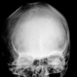
Imaging studies: Imaging studies such as an X-ray can help a clinician assess skeletal abnormalities, such as short, asymmetrical, or poorly developed ribs or rounded shoulders, which may aid in the diagnosis of craniofrontonasal dysplasia.
Genetic testing: If craniofrontonasal dysplasia is suspected, a genetic test may be performed to confirm a diagnosis. A sample of the patient's blood is taken and analyzed in a laboratory for a defect in the EFNB1 gene. If this is detected, a positive diagnosis is made.
Prenatal DNA testing: If there is a family history of craniofrontonasal dysplasia, prenatal testing may be performed to determine whether the fetus has the disorder. Amniocentesis and chorionic villus sampling (CVS) can diagnose the condition. However, because there are serious risks associated with these tests, patients should discuss the potential health benefits and risks with a medical professional.
During amniocentesis, a long, thin needle is inserted through the abdominal wall and into the uterus, and a small amount of amniotic fluid is removed from the sac surrounding the fetus. Cells in the fluid are then analyzed for normal and abnormal chromosomes. This test is performed after 15 weeks of pregnancy. The risk of miscarriage is about one in 200-400 patients. Some patients may experience minor complications, such as cramping, leaking fluid, or irritation where the needle was inserted.
During chorionic villus sampling (CVS), a small piece of tissue (chorionic villi) is removed from the placenta between the ninth and 14th weeks of pregnancy. CVS may be performed through the cervix or through the abdomen. The cells in the tissue sample are then analyzed for the mutation in the EFNB1 gene. Miscarriage occurs in about 0.5-1% of women who undergo this procedure.
Complications
Limited mobility: Problems with the bones of the hands and feet, and with other bones may cause people with craniofrontonasal dysplasia to have limited mobility. This may impair fine motor skills as well as the ability to perform activities of daily living, such as dressing or bathing.
Treatment
General: There is currently no known cure for craniofrontonasal dysplasia. Treatment aims to reduce symptoms and prevent complications.
Corrective lenses: Differences in visual acuity between the right and left eye may be corrected by wearing glasses or contact lenses.
Physical therapy: Patients with craniofrontonasal dysplasia may benefit from seeing a physical therapist, who can help them maintain muscle tone and keep muscles strong and flexible through performing different exercises.
Occupational therapy: Patients with craniofrontonasal dysplasia may benefit from occupational therapy. During sessions, a therapist helps the child learn skills needed to perform basic daily tasks, such as feeding, dressing, and communicating with others. Parents and caregivers can ask their children's pediatricians to recommend a therapist.
Surgery: Individuals with craniofrontonasal dysplasia may require surgery to correct abnormalities of the fingers, toes, some bones, and sometimes even the skull. Depending on the severity of eye problems, surgery may be helpful in correcting crossed eyes (strabismus) and lazy eye (amblyopia).
Integrative Therapies
There is currently a lack of information regarding the use of integrative therapies for the treatment or prevention of craniofrontonasal dysplasia.
Prevention
General: Because craniofrontonasal dysplasia is an inherited condition, there is currently no known way to prevent the disease. However, a number of options are available for patients with family histories of the condition.
Genetic testing and counseling: Individuals who have craniofrontonasal dysplasia may meet with a genetic counselor to discuss the risks of having children with the disease. Genetic counselors can explain the options and the associated risks of various tests, including pre-implantation genetic diagnosis (PGD), amniocentesis, and chorionic villus sampling (CVS).
Pre-implantation genetic diagnosis (PGD) may be used in combination with in vitro (artificial) fertilization. In PGD, embryos are tested for the defective EFNB1 gene, and only the embryos that are not affected are selected for implantation. Because craniofrontonasal dysplasia can be detected in an unborn baby, parents may choose whether to continue the pregnancy. Genetic counselors may assist parents with these difficult decisions.
Author Information
This information has been edited and peer-reviewed by contributors to the Natural Standard Research Collaboration (www.naturalstandard.com).
Bibliography
Natural Standard developed the above evidence-based information based on a thorough systematic review of the available scientific articles. For comprehensive information about alternative and complementary therapies on the professional level, go to www.naturalstandard.com. Selected references are listed below.
FACES: The National Craniofacial Association. www.faces-cranio.org.
Feldman GJ, Ward DE, Lajeunie-Renier E, et al. A novel phenotypic pattern in X-linked inheritance: craniofrontonasal syndrome maps to Xp22. Hum. Molec. Genet. 6: 1937-41, 1997. View Abstract
Forward Face, Inc. www.forwardface.org.
Grutzner E, Gorlin RJ. Craniofrontonasal dysplasia: phenotypic expression in females and males and genetic considerations. Oral Surg. Oral Med. Oral Path. 65: 436-44, 1988. View Abstract
Kapusta L, Brunner HG, Hamel BCJ. Craniofrontonasal dysplasia. Europ. J. Pediat. 151: 837-41, 1992. View Abstract
Michels VV, Derleth DP, Hoffman AD, et al. Craniofrontonasal dysostosis with deafness and axillary pterygia. Am. J. Med. Genet. 34: 445-50, 1989. View Abstract
Morris CA, Palumbos JC, Carey JC. Delineation of the male phenotype in craniofrontonasal syndrome. Am. J. Med. Genet. 27: 623-31, 1987. View Abstract
Natural Standard: The Authority on Integrative Medicine. www.naturalstandard.com.
Saavedra D, Richieri-Costa A, Guion-Almeida ML, et al. Craniofrontonasal syndrome: study of 41 patients. Am. J. Med. Genet. 61: 147-51, 1996. View Abstract
Sax CM, Flannery DB. Craniofrontonasal dysplasia: clinical and genetic analysis. Clin. Genet. 29: 508-15, 1986. View Abstract
Twigg SRF, Kan R, Babbs C, et al. Mutations of ephrin-B1 (EFNB1), a marker of tissue boundary formation, cause craniofrontonasal syndrome. Proc. Nat. Acad. Sci. 101: 8652-7, 2004. View Abstract
Wieland I, Jakubiczka S, Muschke P, et al. Mutations of the ephrin-B1 gene cause craniofrontonasal syndrome. Am. J. Hum. Genet. 74: 1209-15, 2004. View Abstract
Wieland I, Reardon W, Jakubiczka S, et al. Twenty-six novel EFNB1 mutations in familial and sporadic craniofrontonasal syndrome (CFNS). Hum. Mutat. 26: 113-8, 2005. View Abstract
Copyright © 2013 Natural Standard (www.naturalstandard.com)
The information in this monograph is intended for informational purposes only, and is meant to help users better understand health concerns. Information is based on review of scientific research data, historical practice patterns, and clinical experience. This information should not be interpreted as specific medical advice. Users should consult with a qualified healthcare provider for specific questions regarding therapies, diagnosis and/or health conditions, prior to making therapeutic decisions.
Updated:
March 22, 2017